Never-Before-Seen Images of Fetal Brain Activity

Until very recently, obtaining images of fetal brain activity was very complicated. Today, thanks to advanced technology, we can see high-quality images that allow us to better understand certain aspects of our development.
Magnetic Resonance Imaging (MRI) of the fetal brain is a diagnostic method that is complementary to the ultrasounds many mothers undergo for a very specific reason: to obtain a morphological and biometric study of the baby’s brain in order to detect any anomalies.
“Life is fascinating. You just have to know how to look at it the right way.”
-Alexandre Dumas-
These tests usually take place after the 20th week of pregnancy, just when the cerebral corpus callosum has already formed and the diagnostics become even safer. Remember, the fetus is suspended in amniotic fluid, and in liquid the resolution of MRI is poor quality, and any movement makes it impossible to obtain clear data.
This type of prenatal test usually has a success rate of 50% when identifying a problem. But now, this has changed completely. At the moment, we have just taken a giant step forward and already have much more precise algorithms with which to take perfect readings about a baby’s brain activity.
What has been discovered in these first diagnostic tests has been a revolution in the field of perinatal medicine. We’ll explain it to you below.

Brain activity of premature babies
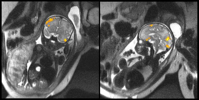
In the image above, we can see the MRI of a fetus at 20 weeks and other of one at 40 weeks. They are images obtained by the faculty of the Wayne State University School of Medicine in Michigan that clearly illustrate the fetal brain activity of these two babies while in their mothers’ wombs.
One of the scientists’ main objectives with this type of test is to study how the neurons of babies are connected during the last weeks of gestation. The data obtained reveal aspects that, until now, were unknown about premature babies.
Low cerebral connectivity in fetuses
Data from this first study have been published in the journal “Scientific Reports.” To carry out this analysis with the new MRIs, 36 pregnant women were followed from week 20 to week 36. Half of them were carrying a high-risk pregnancy and gave birth prematurely.
- It was discovered that the fetuses that would be born before reaching term had a much weaker cerebral connectivity than the other babies in the same week of pregnancy.
- Until now, it was thought that the low cerebral connectivity detected in premature babies was basically due to a traumatic birth or to the hypoxia that babies can end up suffering during childbirth.
However, this new test makes it clear that low neuroconnectivity is already present inside the uterus, and that the scarce connection between neurons is very evident in the Broca area, which is the area responsible for language processing.

How useful are these fetal brain activity tests?
As we pointed out earlier, fetal MRI aims to detect any perinatal anomaly. Today, we cannot ignore the fact that preterm births are becoming more common, a reality that forces doctors, scientists and families themselves to deploy new strategies, energies and resources.
- Data from this study have shown that many of the babies born prematurely had inflamed placental tissue. Something like this makes scientists think that maternal inflammation can determine both the low fetal brain activity and subsequent premature birth.
- Also, the sooner these perinatal anomalies are detected, the greater the chance of intervention. We cannot forget that premature babies are at increased risk of autism, attention deficits and other special learning needs.
When you least expect it, life puts a challenge in front of you.
-Paulo Coelho-

To conclude, these first images about fetal brain activity give us an exceptional window through which we can examine our own development even further. However, it is mainly a precision diagnostic tool that offers more comprehensive attention to premature children, to the lives that are in high need of science, doctors and family.
Until very recently, obtaining images of fetal brain activity was very complicated. Today, thanks to advanced technology, we can see high-quality images that allow us to better understand certain aspects of our development.
Magnetic Resonance Imaging (MRI) of the fetal brain is a diagnostic method that is complementary to the ultrasounds many mothers undergo for a very specific reason: to obtain a morphological and biometric study of the baby’s brain in order to detect any anomalies.
“Life is fascinating. You just have to know how to look at it the right way.”
-Alexandre Dumas-
These tests usually take place after the 20th week of pregnancy, just when the cerebral corpus callosum has already formed and the diagnostics become even safer. Remember, the fetus is suspended in amniotic fluid, and in liquid the resolution of MRI is poor quality, and any movement makes it impossible to obtain clear data.
This type of prenatal test usually has a success rate of 50% when identifying a problem. But now, this has changed completely. At the moment, we have just taken a giant step forward and already have much more precise algorithms with which to take perfect readings about a baby’s brain activity.
What has been discovered in these first diagnostic tests has been a revolution in the field of perinatal medicine. We’ll explain it to you below.

Brain activity of premature babies
In the image above, we can see the MRI of a fetus at 20 weeks and other of one at 40 weeks. They are images obtained by the faculty of the Wayne State University School of Medicine in Michigan that clearly illustrate the fetal brain activity of these two babies while in their mothers’ wombs.
One of the scientists’ main objectives with this type of test is to study how the neurons of babies are connected during the last weeks of gestation. The data obtained reveal aspects that, until now, were unknown about premature babies.
Low cerebral connectivity in fetuses
Data from this first study have been published in the journal “Scientific Reports.” To carry out this analysis with the new MRIs, 36 pregnant women were followed from week 20 to week 36. Half of them were carrying a high-risk pregnancy and gave birth prematurely.
- It was discovered that the fetuses that would be born before reaching term had a much weaker cerebral connectivity than the other babies in the same week of pregnancy.
- Until now, it was thought that the low cerebral connectivity detected in premature babies was basically due to a traumatic birth or to the hypoxia that babies can end up suffering during childbirth.
However, this new test makes it clear that low neuroconnectivity is already present inside the uterus, and that the scarce connection between neurons is very evident in the Broca area, which is the area responsible for language processing.

How useful are these fetal brain activity tests?
As we pointed out earlier, fetal MRI aims to detect any perinatal anomaly. Today, we cannot ignore the fact that preterm births are becoming more common, a reality that forces doctors, scientists and families themselves to deploy new strategies, energies and resources.
- Data from this study have shown that many of the babies born prematurely had inflamed placental tissue. Something like this makes scientists think that maternal inflammation can determine both the low fetal brain activity and subsequent premature birth.
- Also, the sooner these perinatal anomalies are detected, the greater the chance of intervention. We cannot forget that premature babies are at increased risk of autism, attention deficits and other special learning needs.
When you least expect it, life puts a challenge in front of you.
-Paulo Coelho-

To conclude, these first images about fetal brain activity give us an exceptional window through which we can examine our own development even further. However, it is mainly a precision diagnostic tool that offers more comprehensive attention to premature children, to the lives that are in high need of science, doctors and family.
This text is provided for informational purposes only and does not replace consultation with a professional. If in doubt, consult your specialist.


