Basal Ganglia - Anatomy, Physiology, and Function

Written and verified by the psychologist Valeria Sabater
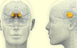
The basal ganglia are a set of groups of neurons specialized in processing information about movement. They’re key when you must use your hands to catch a ball or for moving your feet when you need to climb a ladder or when you drive, paint, or play an instrument, among other activities. These help you fine-tune and adjust every action you carry out throughout the day.
These are a set of subcortical nuclei of the brain that play a very important role. Note, as a curiosity, that they’re not part of the motor system itself. They’re actually a wide set of structures interconnected with different areas. Therefore, their purpose is to plan actions. Especially those with a specific objective that often stem from previous learning.
Neurologists describe the basal ganglia as those neural groups that define the functions of the motor cortex. They ensure, so to speak, the correct execution of any motor plan between the brain and the muscles. Thus, any alteration in these structures irremediably leads to experience alterations in movement. As it happens, for example, in diseases like Parkinson’s.
“The neural processes underlying that which we call creativity have nothing to do with rationality. That is to say, if we look at how the brain generates creativity, we will see that it is not a rational process at all; creativity is not born out of reasoning.”
-Rodolfo R. Llinás-
Basal ganglia, what are they?
The basal ganglia, as you read above, modulates and controls motor activity. However, they carry out many other functions. Thus, studies such as the one conducted by Centro de Investigación Médica Aplicada [Center for Applied Medical Research (CIMA)] at Universidad of Navarra in Spain, indicate the following.
- Firstly, the basal ganglia mediate everything that has to do with motor learning (doing sports, driving, writing, or making crafts). Note that they also have a significant connection to motivation and emotion.
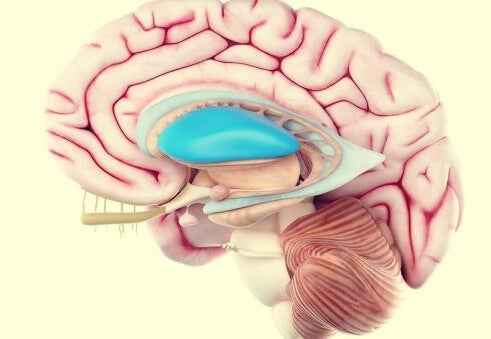
- Similarly, anytime you hear about basal ganglia you must know they’re a series of interconnected subcortical nuclei. Also, they live around the limbic system and the third ventricle (at the level of the temporal lobe).
- The main neurotransmitters that work in these structures are dopamine and GABA.
- Likewise, scientists discovered new data on the anatomy and functional chemistry of the basal ganglia. The kind that helps them better understand its relevance in human behavior and activity. It’s also helpful for treating various diseases.
Composition of the basal ganglia
The various types of basal ganglia are entry nuclei (those that receive information from other brain structures), exit nuclei (those that send information to the thalamus), and intrinsic nuclei (those that act as intermediate zones between the previous ones). Thus, continue reading to find out more about their organization.
Striatum
The striatum is the largest subcortical brain structure in the mammalian brain. It’s an area that acts as an intrinsic nucleus. In other words, it receives information and, in turn, sends it to other areas.
Caudate nucleus
The caudate nucleus is related to the modulation of movement and, also, to various emotional processes. It connects to the frontal lobe and regulates aspects such as motivation, memory, and feelings of threat, among others.
Lentiform nucleus
The lentiform nucleus is a structure that lies just below the caudate nucleus. It’s a fairly large area (about two inches) that facilitates motor skills, regulates your posture and learning processes, and even your motivation.
The putamen nucleus
In recent years, the relationship between the putamen nucleus and feelings of love and hate is under investigation. Likewise, and within the basal ganglia, it performs a basic function in automated movement tasks. It lives next to the caudate nucleus.
The pale balloon
The pale balloon regulates the information established between the putamen nuclei and the caudate towards the thalamus. Likewise, it presents a connection with the limbic system, acting as a reinforcer of behaviors, especially those derived from drug use.
Black matter
The substantia nigra is integrated into the midbrain and is also a key element of the basal ganglia. Its neurons, due to a neuromelanin pigment, give it that dark coloration that tends to increase over the years. It helps you learn, move, and orientate.
In which processes do the basal ganglia mediate?
As you read above, the basal ganglia are made up of various subcortical neuronal structures. They send each other and continuously receive information; thus, participating in processes that go beyond movement.
In fact, studies such as those conducted by doctors Ahmed A. Moustafa and Izhar Bar-Gad, from Bar-Ilan University, Ramat Gan ( אוניברסיטת בר-אילן) in Israel indicate these are key in the processes of memory and motivation. Thus, any alteration is related to conditions such as Parkinson’s disease, attention-deficit/hyperactivity disorder (ADHD), Tourette syndrome, etc.
Therefore, studying disorders such as those mentioned above has made it possible to observe which processes the basal ganglia mediated over the decades. They’re the following:
- Regulation and control of movement.
- Learning-related to procedures that humans end up automating such as driving.
- Activities and movements related to planning.
- Motivational and emotional processes.
Diseases and disorders related to the basal ganglia
Much of the observed abnormalities in the basal ganglia often have serious implications. In fact, many of these diseases that are etiologically related to these structures have a degenerative nature. They’re the following:
- Parkinson’s disease.
- Huntington’s disease.
- PAP syndrome (a clinical condition related to a serious lack of motivation).
- Tourette syndrome.
In conclusion, note that conditions such as obsessive-compulsive disorder and attention deficit disorder – with or without hyperactivity (ADHD) – are also associated with small alterations in the basal ganglia. However, psychological treatments can be very effective in these cases.
The basal ganglia are a set of groups of neurons specialized in processing information about movement. They’re key when you must use your hands to catch a ball or for moving your feet when you need to climb a ladder or when you drive, paint, or play an instrument, among other activities. These help you fine-tune and adjust every action you carry out throughout the day.
These are a set of subcortical nuclei of the brain that play a very important role. Note, as a curiosity, that they’re not part of the motor system itself. They’re actually a wide set of structures interconnected with different areas. Therefore, their purpose is to plan actions. Especially those with a specific objective that often stem from previous learning.
Neurologists describe the basal ganglia as those neural groups that define the functions of the motor cortex. They ensure, so to speak, the correct execution of any motor plan between the brain and the muscles. Thus, any alteration in these structures irremediably leads to experience alterations in movement. As it happens, for example, in diseases like Parkinson’s.
“The neural processes underlying that which we call creativity have nothing to do with rationality. That is to say, if we look at how the brain generates creativity, we will see that it is not a rational process at all; creativity is not born out of reasoning.”
-Rodolfo R. Llinás-
Basal ganglia, what are they?
The basal ganglia, as you read above, modulates and controls motor activity. However, they carry out many other functions. Thus, studies such as the one conducted by Centro de Investigación Médica Aplicada [Center for Applied Medical Research (CIMA)] at Universidad of Navarra in Spain, indicate the following.
- Firstly, the basal ganglia mediate everything that has to do with motor learning (doing sports, driving, writing, or making crafts). Note that they also have a significant connection to motivation and emotion.
- Similarly, anytime you hear about basal ganglia you must know they’re a series of interconnected subcortical nuclei. Also, they live around the limbic system and the third ventricle (at the level of the temporal lobe).
- The main neurotransmitters that work in these structures are dopamine and GABA.
- Likewise, scientists discovered new data on the anatomy and functional chemistry of the basal ganglia. The kind that helps them better understand its relevance in human behavior and activity. It’s also helpful for treating various diseases.
Composition of the basal ganglia
The various types of basal ganglia are entry nuclei (those that receive information from other brain structures), exit nuclei (those that send information to the thalamus), and intrinsic nuclei (those that act as intermediate zones between the previous ones). Thus, continue reading to find out more about their organization.
Striatum
The striatum is the largest subcortical brain structure in the mammalian brain. It’s an area that acts as an intrinsic nucleus. In other words, it receives information and, in turn, sends it to other areas.
Caudate nucleus
The caudate nucleus is related to the modulation of movement and, also, to various emotional processes. It connects to the frontal lobe and regulates aspects such as motivation, memory, and feelings of threat, among others.
Lentiform nucleus
The lentiform nucleus is a structure that lies just below the caudate nucleus. It’s a fairly large area (about two inches) that facilitates motor skills, regulates your posture and learning processes, and even your motivation.
The putamen nucleus
In recent years, the relationship between the putamen nucleus and feelings of love and hate is under investigation. Likewise, and within the basal ganglia, it performs a basic function in automated movement tasks. It lives next to the caudate nucleus.
The pale balloon
The pale balloon regulates the information established between the putamen nuclei and the caudate towards the thalamus. Likewise, it presents a connection with the limbic system, acting as a reinforcer of behaviors, especially those derived from drug use.
Black matter
The substantia nigra is integrated into the midbrain and is also a key element of the basal ganglia. Its neurons, due to a neuromelanin pigment, give it that dark coloration that tends to increase over the years. It helps you learn, move, and orientate.
In which processes do the basal ganglia mediate?
As you read above, the basal ganglia are made up of various subcortical neuronal structures. They send each other and continuously receive information; thus, participating in processes that go beyond movement.
In fact, studies such as those conducted by doctors Ahmed A. Moustafa and Izhar Bar-Gad, from Bar-Ilan University, Ramat Gan ( אוניברסיטת בר-אילן) in Israel indicate these are key in the processes of memory and motivation. Thus, any alteration is related to conditions such as Parkinson’s disease, attention-deficit/hyperactivity disorder (ADHD), Tourette syndrome, etc.
Therefore, studying disorders such as those mentioned above has made it possible to observe which processes the basal ganglia mediated over the decades. They’re the following:
- Regulation and control of movement.
- Learning-related to procedures that humans end up automating such as driving.
- Activities and movements related to planning.
- Motivational and emotional processes.
Diseases and disorders related to the basal ganglia
Much of the observed abnormalities in the basal ganglia often have serious implications. In fact, many of these diseases that are etiologically related to these structures have a degenerative nature. They’re the following:
- Parkinson’s disease.
- Huntington’s disease.
- PAP syndrome (a clinical condition related to a serious lack of motivation).
- Tourette syndrome.
In conclusion, note that conditions such as obsessive-compulsive disorder and attention deficit disorder – with or without hyperactivity (ADHD) – are also associated with small alterations in the basal ganglia. However, psychological treatments can be very effective in these cases.
All cited sources were thoroughly reviewed by our team to ensure their quality, reliability, currency, and validity. The bibliography of this article was considered reliable and of academic or scientific accuracy.
- Alexander, G.E.; DeLong, M.R. & Strick, P.L. (1986). Parallel organization of functionally segregated circuits linking basal ganglia and cortex. Annu Rev Neurosci.; 9:357 – 381.
- Báez-Mendoza, R., & Schultz, W. (2013). The role of the striatum in social behavior. Frontiers in neuroscience, 7, 233.
- Driscoll, M. E., Bollu, P. C., & Tadi, P. (2021). Neuroanatomy, nucleus caudate. StatPearls. https://www.ncbi.nlm.nih.gov/books/NBK557407/
- Ghandili, M., & Munakomi, S. (2021). Neuroanatomy, putamen. StatPearls. https://www.ncbi.nlm.nih.gov/books/NBK542170/
- Graybiel, A. M., & Grafton, S. T. (2015). The striatum: where skills and habits meet. Cold Spring Harbor perspectives in biology, 7(8), a021691.
- Hollerman, J. R., Tremblay, L., & Schultz, W. (2000). Involvement of basal ganglia and orbitofrontal cortex in goal-directed behavior. Progress in brain research, 126, 193-215.
- Kandel, E. R. (2001). Principios de Neurociencia. 1º edición. McGraw-Hill.
- Lanciego, J. L., Luquin, N., & Obeso, J. A. (2012). Functional neuroanatomy of the basal ganglia. Cold Spring Harbor perspectives in medicine, 2(12), a009621.
- Martin, J.H. (1998). Neuroanatomía. Madrid: Prentice Hall.
- Mathai, A., & Smith, Y. (2011). The corticostriatal and corticosubthalamic pathways: two entries, one target. So what?. Frontiers in systems neuroscience, 5, 64.
- Moustafa, A. A., Bar-Gad, I., Korngreen, A., & Bergman, H. (2014). Basal ganglia: physiological, behavioral, and computational studies. Frontiers in systems neuroscience, 8, 150.
- Netter, F.M. (1987). Sistema Nervioso, Anatomía y Fisiología. A Colección Ciba de Ilustraciones Médicas (Vl 1). Barcelona: Salvat.
- Nolte, J. (1994). El cerebro humano: introducción a la anatomía funcional. Madrid: Mosby-Doyma.
- Seger, C. A., & Cincotta, C. M. (2005). The roles of the caudate nucleus in human classification learning. Journal of Neuroscience, 25(11), 2941-2951.
- Sonne, J., Reddy, V., & Beato, M. R. (2019). Neuroanatomy, substantia nigra. StatPearls. https://www.ncbi.nlm.nih.gov/books/NBK536995/
- White, N. M. (2009). Some highlights of research on the effects of caudate nucleus lesions over the past 200 years. Behavioural brain research, 199(1), 3-23.
- Young, C. B., Reddy, V., & Sonne, J. (2021). Neuroanatomy, basal ganglia. https://www.ncbi.nlm.nih.gov/books/NBK537141/#:~:text=The%20basal%20ganglia’s%20primary%20function,what%20actions%20to%20%E2%80%9Cdisinhibit%E2%80%9D.
This text is provided for informational purposes only and does not replace consultation with a professional. If in doubt, consult your specialist.