Hippocampal Formation: Structure and Functions
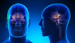
Cognitive processes, such as learning and memory, are crucial to human beings. The hippocampus plays a fundamental role in these processes, which is one of the hippocampal formation regions.
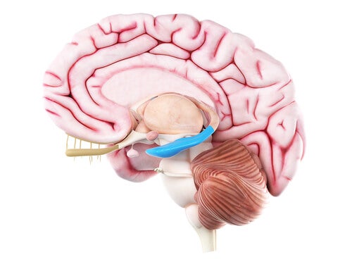
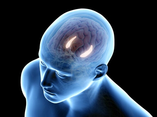
Hippocampal formation is a C-shaped prominent structure, located in the lower part of a lateral ventricle in the brain. The hippocampus itself consists of three main subfields (CA1 -CA3). But is hippocampal formation just the hippocampus?
Anatomic analysis of hippocampal formation
In the 16th century, anatomist Arantius (2) mentioned hippocampal formation for the first time. He named it hippocampus, which comes from the Greek word for seahorses.
However, hippocampal formation isn’t just the hippocampus. In fact, it also consists of the dentate gyrus, the subicular complex, and the entorhinal cortex.
Thus, hippocampal formation is about five centimeters long. In the middle part, there’s the uncus as well, which is potato-shaped and varies a lot from brain to brain.
Taking into account its position from the corpus callosum, the hippocampus has three parts: precommisural, supracommisural, and retrocommisural.
Hippocampal formation: Architecture
Dentate gyrus
The dentate gyrus is the most medial part of the cerebral cortex. When it comes to cytoarchitectonics, the dentate gyrus is a trilaminate cortical area. In hippocampal formation, the dentate gyrus, in its typical C-shape, is ventrally separated from the first hippocampus part and from the subiculum at the hippocampus fissure.
This structure’s main cell layer is full of granular cell bodies. The apical dendrites of these cells have ramifications in the dentate molecular layer. The granular cells and the molecular layers combined represent the fascia dentata.
Then, the third innermost layer of the dentate gyrus is the polymorphic layer or hilus. Next to it, there’s a fraction of the pyramidal cell layer enclosed by the granular cells.
The hippocampus
The hippocampus has subfields named CA1, CA2, and CA3 that consist of one cell layer: the pyramidal cell layer. The surface that confines with the ventricular lumen, formed by axons of pyramidal cells, is called alveus. Historically, this region consists of:
- Lucidum stratum
- Radiatum stratum
- Lacunosum-moleculare
The lucidum stratum, CA3, has fibers that form proximal dendritic synapses above the pyramidal cell wall of this layer. CA2 layer is relatively compact and presents a pyramidal cell layer as well, but its edges are hard to define.
Furthermore, the CA1 layer is a hippocampus subfield (3). The pyramidal cell layer in this region consists, in turn, of one external and one internal layer.
Subiculum
The CA1 layer and the subiculum overlap in the edges, forming a transition area. The subiculum is divided primarily in the following layers:
- Superficially, there’s a wide molecular layer where subicular pyramidal-cell dendrites are located. In turn, this pyramidal cell layer can be divided into two sub-layers: external and internal.
- External layer cells have a lipofuscin pigment accumulation in its apical dendrites.
- The presubiculum consists of a superficial layer, containing modified pyramidal neurons.
- The parasubiculum has one cell layer that’s hard to differentiate from the presubiculum.
Entorhinal cortex
The term ‘entorhinal cortex’ is a synonym of the Brodmann area. It mainly extends rostrally toward the middle part of the amygdala, and caudally toward the anterior commissure of the lateral geniculate nucleus.
This area is somewhat different from the rest of the brain regions.
Hippocampal formation connectivity
Hippocampal intrinsic circuit
Connectivity of the hippocampal information follows a unidirectional and glutamatergic (excitatory) path that’s part of a closed circuit. In this intrinsic chain of connections, the dentate gyrus is very important, since it receives the most information which is then transmitted to the entorhinal cortex.
Extrinsic connections
The hippocampal extrinsic circuit is formed by:
- Several cortical arteries.
- The amygdaloid complex.
- The medial septal nucleus.
- Thalamus.
- Supramammilary nucleus.
- The monoaminergic nucleus of the brainstem.
That’s apparently how the hippocampus receives sensory information from a variety of cortical regions.
Cortical connections
These projections serve mainly to introduce sensory information into the hippocampal formation.
Subcortical connections
The fimbriae and the fornix form the classical efferent system of hippocampal formation. In addition, there are also great connections between hippocampal formation and the amygdala.
Lastly, the connections produced between the hippocampal formation and the hypothalamus establish through the subiculum.
As you can see, the hippocampal formation is a complex set of regions that include the hypothalamus. Although most research projects have studied animals, it seems clear that the regions here described are very similar to human hippocampal formation.
All cited sources were thoroughly reviewed by our team to ensure their quality, reliability, currency, and validity. The bibliography of this article was considered reliable and of academic or scientific accuracy.
-
Insausti, R., & Amaral, D. G. (2003). Hippocampal formation. In The Human Nervous System: Second Edition. Elsevier Inc..
- Arantius G (1587). De humano foetu. Ejusdem anatomicorum observationum liber, etc. Venice, pp 44–45.
-
Stephan, H. (1983). Evolutionary trends in limbic structures. Neuroscience & Biobehavioral Reviews, 7(3), 367-374.
This text is provided for informational purposes only and does not replace consultation with a professional. If in doubt, consult your specialist.