Biopsychology Research Methods
Biopsychology research methods have evolved in the last decades. Although there are several biopsychology research methods, in this article, we’ll focus on those that study what happens in the brain under certain conditions.
Authors such as Dewsbury (1991) define biopsychology as “the scientific study of the biology of behavior”, a field also called psychobiology. However, other authors prefer the term biopsychology because it “indicates a biological approach to the study of psychology, more than a psychological approach to the study of biology.”
Human brain stimulation and visualization methods
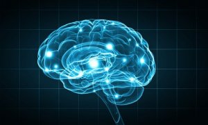
Observing and recording brain activity is a very important milestone that was achieved thanks to the different techniques that scientists developed during the 20th century. These biopsychology research methods are, without a doubt, a breakthrough in the study of our most curious organ.
Contrast radiography
This technique consists of injecting a substance in the body to absorb X-rays. This way, scientists see the contrast between the compartment and the tissue around it.
Cerebral angiography is a kind of contrast radiography. To do it, a contrast medium is inserted into a brain vessel. The aim is to observe the circulatory system while performing an X-ray. This technique is very useful to locate vessel injuries and brain tumors.
Computerized axial tomography scan (CT scan)
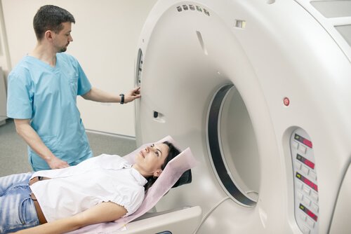
Through a CT scan, experts can see the whole brain structure. During the test, the patient lies down in the middle of a big cylinder. While the patient holds still, an X-ray tube and a receptor take many separate photographs. This happens while the emitter and the receptor spin around the patient’s head.
All this information is transmitted to a computer, which allows doctors to explore the brain on a horizontal plane. Usually, they do it on eight to nine horizontal brain sections. Once all explorations are combined, it’s possible to make a three-dimensional representation of the brain.
Nuclear magnetic resonance (NMR)
NMR facilitates high-resolution imaging thanks to the different waves hydrogen atoms emit when activated by radio frequency on a magnetic field. It provides a high spatial resolution and produces three-dimensional images.
Positron emission tomography (PET)
PET provides brain activity images instead of brain structure images. To get the images, scientists inject radioactive fludeoxyglucose (FDG) in the carotid artery. Active neurons rapidly absorb FDG, which accumulates once neurons don’t metabolize it anymore, then it slowly degrades. This is how one can observe which neurons are active at a given time during different activities.
Functional magnetic resonance imaging (fMRI)
On the other hand, MRIs offer an image of the increase in the amount of oxygen present in the brain’s blood. Thus, it successfully measures brain activity. If we compare it to the PET, it actually has four advantages:
- Doctors don’t have to inject the patient with anything.
- It provides both functional and structural information.
- It offers a better spatial resolution.
- Provides three-dimensional images of the entire brain.
Magnetoencephalography (MEG)
It measures the changes in the magnetic fields located on the scalp’s surface. These changes occur due to the variations in the neuronal activity guidelines.
Transcranial magnetic stimulation (TMS)
Walsh and Rothwell (2000) state that TMS “alters an area of the cortex, creating a magnetic field under the coil that goes over the cranium.” TMS basically “turns off” a part of the brain temporarily, to study behavior and cognition under those circumstances.
Lesion methods
Lesion methods focus on the destruction of a small brain area to see how it affects behavior.
- Aspiration lesion. This method creates a lesion in an exposed or easily accessible cortical tissue area. The doctors remove the tissue with a fine-point crystal pipette.
- Radio-frequency lesion. It’s carried out by creating small subcortical lesions. For that, an electrode channels the high-frequency current through the tissue of interest. The size and shape of the lesion depend on three factors:
- Duration of the procedure.
- Intensity of the current.
- Configuration of the electrode’s point.
- Scalpel cuts. It consists of sectioning the brain area of interest.
- Cooling lesion. This biopsychology research method, although included under lesion methods, is actually temporary and reversible. Instead of destroying tissue, one area is cooled a bit over the freezing point. Neurons stop emitting signals, so the cold brain area remains blocked. With this, researchers are able to see what behavior alterations are caused by those areas. Once the temperature goes back to normal, brain function is restored.
Electrical stimulation
Another biopsychology research method is electrical stimulation. The procedure consists of electrically stimulating a nervous system structure to get data about its functions. Usually, a bipolar electrode is used.
This stimulation “shoots” neurons and alters their behavior. In general, it tends to get the opposite effect of lesion methods. For example, if it’s possible to drastically reduce sleep hours with a lesion, then sleep behavior can become inconvenient and harder to control with electrical stimulation.
Lesion methods with electrical recording
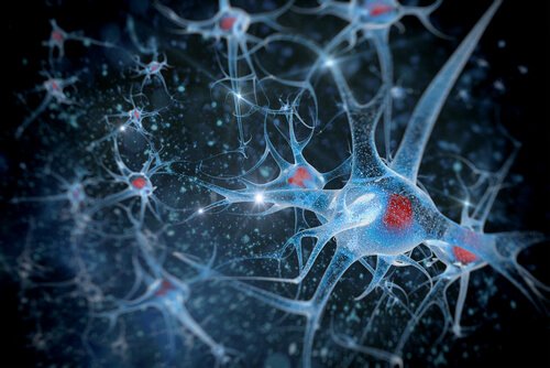
- Intracellular recording. This technique is carried out by introducing a microelectrode in the interior of a neuron. It records fluctuations of membrane potential.
- Extracellular unit recording. A microelectrode is placed in the extracellular fluid that surrounds the neuron. It doesn’t provide information on membrane potential.
- Multi-unit recording. In this case, the electrode point is bigger than that of a microelectrode, so it captures the signals of many neurons at the same time. The detected potentials then go on a circuit that integrates them.
- Invasive EEG monitoring. The stainless steel electrodes go inside the cranium. For subcortical signals, the usual electrodes are made of cable and implanted through stereotactic surgery.
“Anthropology, biology, physiology and psychology have accumulated mountains of material to raise up before mankind in their full scope the tasks of perfecting and developing body and spirit.”
-Leon Trotsky-
Biopsychology research methods: A long way to go
In this article, we talked about the most important biopsychology research methods. However, it’s also worth mentioning that there are other biopsychology research methods that study other body areas, such as muscle tension measurements, eye movement recordings, skin conductance, or cardiovascular activity.
Without a doubt, the breakthrough in this field has been spectacular, but not conclusive. Perhaps in a few years, scientists will come up with new techniques that will contribute to the evolution of neuroscience, which will help improve the quality of life of many people affected by neuronal alterations.
Biopsychology research methods have evolved in the last decades. Although there are several biopsychology research methods, in this article, we’ll focus on those that study what happens in the brain under certain conditions.
Authors such as Dewsbury (1991) define biopsychology as “the scientific study of the biology of behavior”, a field also called psychobiology. However, other authors prefer the term biopsychology because it “indicates a biological approach to the study of psychology, more than a psychological approach to the study of biology.”
Human brain stimulation and visualization methods
Observing and recording brain activity is a very important milestone that was achieved thanks to the different techniques that scientists developed during the 20th century. These biopsychology research methods are, without a doubt, a breakthrough in the study of our most curious organ.
Contrast radiography
This technique consists of injecting a substance in the body to absorb X-rays. This way, scientists see the contrast between the compartment and the tissue around it.
Cerebral angiography is a kind of contrast radiography. To do it, a contrast medium is inserted into a brain vessel. The aim is to observe the circulatory system while performing an X-ray. This technique is very useful to locate vessel injuries and brain tumors.
Computerized axial tomography scan (CT scan)
Through a CT scan, experts can see the whole brain structure. During the test, the patient lies down in the middle of a big cylinder. While the patient holds still, an X-ray tube and a receptor take many separate photographs. This happens while the emitter and the receptor spin around the patient’s head.
All this information is transmitted to a computer, which allows doctors to explore the brain on a horizontal plane. Usually, they do it on eight to nine horizontal brain sections. Once all explorations are combined, it’s possible to make a three-dimensional representation of the brain.
Nuclear magnetic resonance (NMR)
NMR facilitates high-resolution imaging thanks to the different waves hydrogen atoms emit when activated by radio frequency on a magnetic field. It provides a high spatial resolution and produces three-dimensional images.
Positron emission tomography (PET)
PET provides brain activity images instead of brain structure images. To get the images, scientists inject radioactive fludeoxyglucose (FDG) in the carotid artery. Active neurons rapidly absorb FDG, which accumulates once neurons don’t metabolize it anymore, then it slowly degrades. This is how one can observe which neurons are active at a given time during different activities.
Functional magnetic resonance imaging (fMRI)
On the other hand, MRIs offer an image of the increase in the amount of oxygen present in the brain’s blood. Thus, it successfully measures brain activity. If we compare it to the PET, it actually has four advantages:
- Doctors don’t have to inject the patient with anything.
- It provides both functional and structural information.
- It offers a better spatial resolution.
- Provides three-dimensional images of the entire brain.
Magnetoencephalography (MEG)
It measures the changes in the magnetic fields located on the scalp’s surface. These changes occur due to the variations in the neuronal activity guidelines.
Transcranial magnetic stimulation (TMS)
Walsh and Rothwell (2000) state that TMS “alters an area of the cortex, creating a magnetic field under the coil that goes over the cranium.” TMS basically “turns off” a part of the brain temporarily, to study behavior and cognition under those circumstances.
Lesion methods
Lesion methods focus on the destruction of a small brain area to see how it affects behavior.
- Aspiration lesion. This method creates a lesion in an exposed or easily accessible cortical tissue area. The doctors remove the tissue with a fine-point crystal pipette.
- Radio-frequency lesion. It’s carried out by creating small subcortical lesions. For that, an electrode channels the high-frequency current through the tissue of interest. The size and shape of the lesion depend on three factors:
- Duration of the procedure.
- Intensity of the current.
- Configuration of the electrode’s point.
- Scalpel cuts. It consists of sectioning the brain area of interest.
- Cooling lesion. This biopsychology research method, although included under lesion methods, is actually temporary and reversible. Instead of destroying tissue, one area is cooled a bit over the freezing point. Neurons stop emitting signals, so the cold brain area remains blocked. With this, researchers are able to see what behavior alterations are caused by those areas. Once the temperature goes back to normal, brain function is restored.
Electrical stimulation
Another biopsychology research method is electrical stimulation. The procedure consists of electrically stimulating a nervous system structure to get data about its functions. Usually, a bipolar electrode is used.
This stimulation “shoots” neurons and alters their behavior. In general, it tends to get the opposite effect of lesion methods. For example, if it’s possible to drastically reduce sleep hours with a lesion, then sleep behavior can become inconvenient and harder to control with electrical stimulation.
Lesion methods with electrical recording
- Intracellular recording. This technique is carried out by introducing a microelectrode in the interior of a neuron. It records fluctuations of membrane potential.
- Extracellular unit recording. A microelectrode is placed in the extracellular fluid that surrounds the neuron. It doesn’t provide information on membrane potential.
- Multi-unit recording. In this case, the electrode point is bigger than that of a microelectrode, so it captures the signals of many neurons at the same time. The detected potentials then go on a circuit that integrates them.
- Invasive EEG monitoring. The stainless steel electrodes go inside the cranium. For subcortical signals, the usual electrodes are made of cable and implanted through stereotactic surgery.
“Anthropology, biology, physiology and psychology have accumulated mountains of material to raise up before mankind in their full scope the tasks of perfecting and developing body and spirit.”
-Leon Trotsky-
Biopsychology research methods: A long way to go
In this article, we talked about the most important biopsychology research methods. However, it’s also worth mentioning that there are other biopsychology research methods that study other body areas, such as muscle tension measurements, eye movement recordings, skin conductance, or cardiovascular activity.
Without a doubt, the breakthrough in this field has been spectacular, but not conclusive. Perhaps in a few years, scientists will come up with new techniques that will contribute to the evolution of neuroscience, which will help improve the quality of life of many people affected by neuronal alterations.
This text is provided for informational purposes only and does not replace consultation with a professional. If in doubt, consult your specialist.