Anatomy of the Encephalon
This article is about the anatomy of the encephalon, the upper part of the central nervous system. This organ is inside the skull and its purpose is to conduct and integrate information, reasoning, judgment and control behavior. As for its physiological distribution, you can divide it into three distinct parts: the forebrain, the midbrain, and the hindbrain.
Each one of these brain regions contains specific parts that perform different activities. Of all the functions the encephalon performs, the main one is to control body activity and the reception of internal and external information.
Thus, the brain is responsible for associating physical components with psychological ones. Also, it’s responsible for adapting the brain information with what comes from the outside through the senses.
The encephalon is a very wide region. In fact, it’s the most voluminous structure of the human brain. For this reason, it contains thousands of different parts.
Anatomy of the encephalon – the forebrain
The forebrain is the anterior portion of the brain. Thus, it’s one of the first regions to develop during the gestation of an embryo. Subsequently, there are two regions that cover its structure within the forebrain: the telencephalon and the diencephalon.
Telencephalon
The telencephalon is the upper and most bulky region of the forebrain. Thus, it represents the highest level of somatic and vegetative integration.
This region is different between amphibians and mammals. The first one is made of highly developed olfactory bulbs, while the latter has two cerebral hemispheres. Also, the following lobes are inside the telencephalon:
- Occipital. It performs visual sensory operations.
- Parietal. This one processes sensitive and kinesic information.
- Temporary. It performs auditory processes.
- Frontal. This one performs superior functions such as judgment, reasoning, perception and motor control.
- Striated body. It receives information from the cerebral cortex and basal ganglia.
- Rhinocephalus. This is the brain region involved in smell.
Thus, the telencephalon contains multiple brain regions and performs multiple mental processes. Thus, the processing of information from the senses and other brain regions are the most important. However, it also participates in more elaborate functions through the frontal lobe.
Diencephalon
The diencephalon is the other subregion of the forebrain. It’s located under the telencephalon and borders the lower part with the midbrain.
This structure contains very important brain elements. The main ones are the thalamus and the hypothalamus.
- Hypothalamus. This is a small organ and the base of the thalamus that controls autonomous visceral functions and sexual impulses. Also, it performs important activities in the regulation of appetite, thirst, and sleep.
- Thalamus. This is the most voluminous and important region of the diencephalon. In fact, its main function is to collect information from all the senses, except for smell. It’s directly connected to the cerebral cortex and plays an important role in the development of emotions and feelings.
- Subthalamus. This is a small region between the thalamus and the hypothalamus. Thus, it receives information from the cerebellum and the red nucleus and is mainly composed of gray matter.
- Epithalamus. This one is above the thalamus, made up of the pineal gland and the habenular nuclei. The epithalamus belongs to the limbic system and is responsible for producing melatonin.
- Metathalamus. This is above the epithalamus and it’s a structure that acts as a passageway for nerve impulses. These, in turn, circulate from the lower peduncle to the auditory cortex.
Third ventricle
Finally, there’s a ventricle that’s responsible for cushioning craniocephalic strokes in the upper part of the diencephalon to protect the lower regions of the diencephalon.
Anatomy of the encephalon – the midbrain
The midbrain or middle brain is the central part of the brain. Thus, it constitutes the upper structure of the brain stem and is responsible for joining the Varolium bridge and the cerebellum with the diencephalon. Also, there are three main regions within the midbrain:
- Previous. The Tuber cinereum is in this region together with the subsequent perforated substance. What this means is it has a small groove that has its origin in the ocular motor nerve.
- Lateral. This one is made up of the upper conjunctival arm and the optical belt. Thus, their functions are to serve as connections between the tubers and the geniculate bodies.
- Subsequent. It houses the corpora quadrigemina, which are rounded eminences divided into anterior and superior pairs that modulate the visual reflexes. Also, the posterior and inferior ones that modulate the auditory reflexes.
The main function of the midbrain is to drive the motor impulses from the cerebral cortex to the brainstem bridge or from the upper regions of the brain to the lower regions so that they reach the muscles. Thus, it mainly transmits sensory impulses and reflexes and connects the spinal cord with the thalamus.
Anatomy of the encephalon: the hindbrain or rhombencephalon
The rhombencephalon or hindbrain is the lower portion of the brain. It surrounds the fourth cerebral ventricle and borders the lower part with the spinal cord. In fact, it’s made up of the metencephalon, which contains the cerebellum and the bump, and the myelencephalon, which contains the medulla oblongata.
Metencephalon
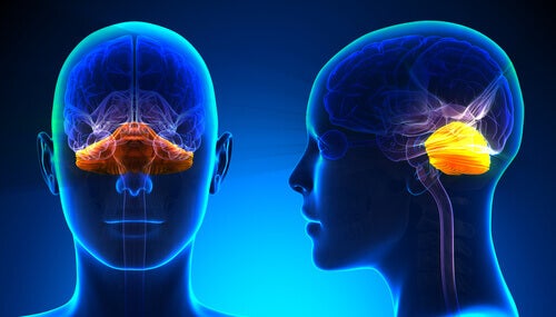
This is the second vesicle of the brain and configures the upper part of the rhombencephalon. Also, it contains two main and important regions for brain functioning: the cerebellum and the bump.
- Cerebellum. Its main function tries to integrate the sensory and motor pathways. Also, it’s a region full of nerve connections that allows establishing a connection with the spinal cord and with the upper parts of the brain.
- Extrusion. This is the portion of the brain stem that lives between the medulla and the midbrain. Thus, its main function is similar to that of the cerebellum and is also responsible for connecting the midbrain with the upper hemispheres of the brain.
Myelencephalon
The myelencephalon is the lower part of the rhombencephalon. In addition, this region contains the medulla, a cone-shaped structure that transmits the impulses from the spinal cord to the brain.
This text is provided for informational purposes only and does not replace consultation with a professional. If in doubt, consult your specialist.