Why Does the Brain Have Folds?

Throughout humankind’s history, humans have always wanted to know more about the brain and its parts. It’s interesting to know what each side of the brain does. Today, the brain is as interesting as ever. But why does the brain have folds?
Judging by the zoological scale, the link between neurons and learning abilities is lineal. The larger your brain surface, the better you learn. Humans have the largest brain surface and the brain has folds thanks to the cerebral gyrus. In fact, it takes up the most space in a small area.
Animals without gyruses are called lissencephalic and have little chance to learn, while the animals that do have gyruses are called gyrencephala and have more abilities to learn. The human being is the most gyrencephal animal. The human brain has folds, more than any other species, followed by giant apes.
“Language and hearing are seated in the cerebral cortex, the folded gray matter that covers the first couple of millimetres of the outer brain like wrapping paper.”
-Michael Finkel-

Cerebral anatomy: the differences between humans and chimpanzees
In May 2009, Scientific American featured an article by Katherine S. Pollard, a biostatistician from the University of California. She developed a computer program to compare the differences between humans and chimpanzees. The sequence with the biggest differences has 118 nucleotides, which she called HAR1, human accelerated region.
Apparently, HAR1 happens in the human brain, as well as in the brains of other vertebrates. Well, this region of the brain slowly evolved in non-humans. There are just two differences between chickens’ and chimpanzees’ sequences. The differences between chimpanzees and humans are 18.
Testing using culture proves that HAR1 modulates genetic expression and it’s active in neurons involved in the development of the brain cortex. In fact, if the HAR1 activates in damaged cells, the brain develops abnormally and the brain cortex looks different.
An anatomical characteristic of intelligence isn’t just how heavy the brain is but that it has folds.
“The mind that opens up to a new idea never returns to its original size.”
-Albert Einstein-

How does the brain get its folds?
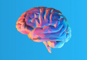
One of the most important characteristics of the human brain is the size of the brain cortex and its folds that look like bumps and grooves on the outside.
Most animals with a big brains have wrinkled brains, while most animals with small brains have a wrinkle-free cortexes.
The neurons are on the top of the brain cortex while, on the bottom, you can find the cable that connects the neurons with the rest of the brain.
In big brains, this neural tissue layer that covers the outside of the brain is larger than the brain structure it covers. Instead of working as a balloon, it folds on itself, making the brain and skull shrink in volume.
Victor Borrel and his team have studied this line and have proved that the radial glial cell, or bRG, plays an important role in expanding the brain cortex.
Thus, the bRG is an important requirement, although not enough by itself, to create a cortex with gyrus because it creates new radial process where neurons migrate. This process expands the brain cortex.
“The human brain starts working the moment you are born and never stops until you stand up to speak in public.”
-George Jessel-

What happens when the brain doesn’t have enough folds?
Cortical folding happens in the womb. The wrinkles in the brain happen around the 20th week of pregnancy and are complete when the child’s one and a half years old.
It’s important to improve the brain’s functions and connections. Besides, it allows adjusting a large cortex into a small cranial space.
Some common pathologies include polymicrogyria, the abnormal development of too many and too small folds, where the neurons ectopically accumulate around the lateral ventricles, forming nodules that can cause epileptic episodes.
All cited sources were thoroughly reviewed by our team to ensure their quality, reliability, currency, and validity. The bibliography of this article was considered reliable and of academic or scientific accuracy.
Del Toro D., Ruff T., Cederfjäll E., Villalba A., Seyit-Bremer G., Borrell V., Klein R. ( 2017 ).” Regulation of cerebral cortex folding by controlling neuronal migration via FLRT adhesion molecules. “ Cell . 169 , 621 – 635.
Rodríguez, O. (2009). ¿Qué región del genoma humano nos distingue de los chimpancés?
Rojo, J. M. I. (1999). La patología cerebral y el conocimiento de nuestra mente. GENES, CULTURA Y MENTE, 97.
Fernández V., Llinares-Benadero C., Borrell V. ( 2016 ). ” Cerebral cortex expansion and folding: what have we learned? “EMBO J . 35 , 1021 – 1044.
This text is provided for informational purposes only and does not replace consultation with a professional. If in doubt, consult your specialist.


