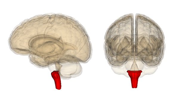
The Spinal Bulb: Structure and Functions
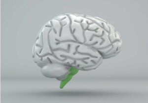
The spinal bulb, or medulla oblongata, is a subcortical structure located in the lower part of the brain stem. In it, one can find nerve connections that carry out sensory and motor functions. What exactly does it do? Why is it so important?
Throughout this article, we’ll answer these questions and explain some curious facts about a brain structure that’s very important for our survival.
The structure of the spinal bulb
The spinal bulb is the lowest structure in the brain stem. Its shape is similar to a cone and it connects the brain to the spinal cord. However, both in composition and function, it isn’t homogeneous. It’s formed by different nuclei, in which we can find important structures such as the following:
- Bulbar pyramids. These are found at the base of the bulb. Here, we can highlight the bundles of nerve fibers that connect the bulb with the cortex and the spine. This is where the pyramidal decussation takes place – especially in the motor pathways. What this means is that there’s a crossing of the nerve fibers from one side to the other. This explains why the cerebral cortex is in charge of controlling the movements of the opposite side of the body.
- Lower cerebellar peduncles. These are located in the posterolateral area. They connect the cerebellum with the upper part of the spinal cord. Nerve fibers pass through them.
- Lemnisks. These are bundles of nerve fibers that transmit information between the brain and the spinal cord. The bundles of lemniscatic fibers also cross, but, in their case, they carry sensory information.
Other elements
- Medial lemnisciae. This is an elongated, thin, highly myelinated structure found on either side of the midline of the spinal cord. It transports information originating from the gracile and cuneiform nuclei. It’s located behind the cerebrospinal fibers and between the olivary bodies (olives).
- Medial longitudinal fascicle. This is a region located next to each medial lemnisciae. It has numerous ascending and descending fibers. Its function is vital for changes in head position and for the coordination of eye movements.
- Olivary body complex. This is located in the trunk of the brain, one part in the cerebral protuberance and another in the spinal cord. It’s a set of oval-shaped nuclei, similar in shape and size to an olive. They regulate voluntary muscle movements.
Nuclei of the spinal bulb
The nuclei consist of clearly differentiated masses of neurons. Let’s have a look at some of them:
- Trigeminal nucleus. In charge of transmitting information related to pain, temperature, and touch.
- Dorsal nucleus of the vagus. This passes through the vagus nerve, hence its name. They’re neural networks that control the enteric nervous system.
- Ambiguous nucleus. The vagus, accessory, and glossopharyngeal nerves belong to, and initiate in, this nucleus. They’re in charge of the muscles of the pharynx and larynx.
- Nucleus of the solitary tract. In charge of controlling visceral sensitivity. It also participates in taste perception.
Functions of the spinal bulb
The spinal bulb is involved in several functions, which are all vital to the body. In fact, if this structure is irretrievably damaged or ceases to function, it leads to death. Let’s have a closer look:
Control of autonomous functions
The spinal bulb is responsible for controlling the involuntary functions of the viscera and maintaining homeostasis. For example:
- Cardiovascular system. It’s in charge of maintaining blood pressure and also regulating the heart rate and maintaining vasoconstriction.
- Breathing. The spinal bulb is in charge of regulating breathing; it manages respiratory function.
- Digestion. It controls the involuntary muscles that are involved in this process. It also regulates the secretion of digestive juices and participates in swallowing processes.
In addition, the spinal bulb manages other involuntary actions:
- Coughing.
- Vomiting.
- Sneezing.

Sensory control
The spinal bulb is also responsible for the transfer of sensory information between the peripheral system and the central nervous system. To do this, it connects the two systems and sends information to the thalamus, which is then communicated to the rest of the brain.
Problems associated with the spinal bulb
If certain cranial pairs (IX, X, XI, XII) are damaged in the bulb, this can cause problems in the regions and functions they control. Here’s a summary of these cranial pairs:
- Glossopharyngeal or Cranial Pair IX. This collects taste and sensory information from the pharynx. It facilitates swallowing by coordinating various neck muscles. In addition, it transmits signals to the salivary gland.
- Vagus nerve or cranial nerve X. Also known as pneumogastric. It innervates the pharynx, esophagus, trachea, bronchi, heart, stomach, and liver. This means that it regulates our autonomic system.
- Accessory nerve or cranial nerve XI. Also known as the spinal nerve. It intervenes in movements of the head, neck, and shoulders.
- Hypoglossal nerve or cranial pair XII. This intervenes in the functioning of the tongue muscles and in swallowing.
The consequences
If one of these fails, then one could experience problems swallowing and even in certain movements. As a result, when this structure doesn’t function properly, then a person could experience:
- Difficulty or paralysis of movement.
- Breathing and heart problems.
- Vertigo.
- Difficulty swallowing.
- Loss of consciousness.
- Weakness.
- Sleepiness.
- Visual and auditory problems.
As you can see, the spinal bulb is absolutely vital for our survival. It’s only just over an inch long and just under an inch wide, but we certainly can’t underestimate it!
All cited sources were thoroughly reviewed by our team to ensure their quality, reliability, currency, and validity. The bibliography of this article was considered reliable and of academic or scientific accuracy.
- Bear, M. F. Connors, B. W., Paradiso, M.A., Nuin, X. U., Guillén, X. V. & Sol Jaquotot, M. J. (2008). Neurociencias: la exploración del cerebro. Wolters Kluwer/Lippincott Williams & Wikins
- De Faes, G. M., Moyano, B.S., Extremera, V.C., Vinues, B.M., Fernández, M. G, Jiménez, M.P., & Rojas, M. R.F. (2018). Revista de neurología, 67(10), 382-386.
- Kandel, E.R; Schwartz, J.H. & Jessell, T.M. (2001). Principios de neurociencia. Madrid: McGrawHill Interamericana.
This text is provided for informational purposes only and does not replace consultation with a professional. If in doubt, consult your specialist.