The Hindbrain: Structure and Functions
Throughout history, scientists have categorized the brain into several parts to try and better understand its functionality and development. One of those parts is the hindbrain, or rhombencephalon, a region that comes from the caudal primary embryonic vesicle.
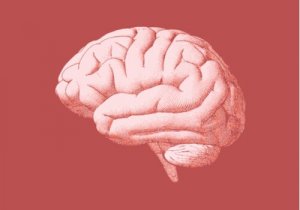
The hindbrain is the brain’s rearmost region. It’s a structure formed by different sub-structures in charge of several essential functions.
Throughout this article, we’ll show you the structure, differentiation process, and functions of this part of the brain.

Hindbrain differentiation process
First of all, you must understand the hindbrain’s origin. For that, it’s important to clarify what exactly a differentiation process is. According to Bear, Connors, and Paradiso, authors of the book Neuroscience: Exploring the Brain, it’s a process through which structures become more complex and functionally specialized.
The first step in the brain differentiation process is the development of three primary brain vesicles of the neural tube, which originate at the rostral extreme.
The most rostral part of the primary vesicles is the prosencephalon, or more commonly known as the forebrain. Then, there’s the vesicle located behind the forebrain, called the mesencephalon or midbrain. And lastly, there’s the most caudal part of the vesicles, which is the hindbrain or rhombencephalon, that connects with the neural tube’s caudal part.
Thus, the hindbrain forms during embryonic development through transversal swellings called rhombomeres, which are basically compartments that allow cells to form groups, each developing in a different way. In addition, they process different functions. The hindbrain has three essential structures:
- Cerebellum. It joins the brainstem in the pons, and it’s a movement-control center fundamental to the organism. It derives from the rostral portion.
- Pons. It’s part of the rostral hindbrain, anterior to the cerebellum and the fourth ventricle.
- Medulla oblongata. Located in a caudal position to the pons and the cerebellum. It derives from the caudal portion.
Also, in the vesicle stage, the rostral hindbrain is shaped like a tube transversely. In the back, the rhombic lip, or the tube’s dorsal-wall tissue, grows rostral and medial-wise until it merges with the opposite side. Furthermore, the crease that forms grows to form the cerebellum. And the tube’s ventral-wall dilates to form the pons.
Moreover, in the differentiation of the hindbrain’s caudal half, some changes occur in the medulla oblongata. On one level, the walls dilate and leave only the “roof” covered in ependymal cells instead of neurons. On another level, in the medulla oblongata, along each side’s ventral surface, there are white matter systems.
Finally, regarding the space for the cerebrospinal fluid, it turns into the fourth ventricle, which continues the mesencephalon’s cerebral aqueduct.
Hindbrain functions
The hindbrain carries out several functions:
- It’s a fundamental part of the brain for information to go through, from the prosencephalon to the bone marrow and vice versa.
- Its neurons play a part in the processing of sensory information.
- The hindbrain’s neurons contribute to voluntary movement control. Plus, they help regulate the autonomic nervous system.
- The cerebellum, which means “little brain”, regulates movement as if it were a control center. It also receives massive axon entries, coming from the bone marrow and the pons. On the other hand, the cerebellum is in charge of comparing the incoming information and calculates the sequences of muscle contractions, which are essential to carry out a movement.
- The medulla oblongata is in charge of carrying the somatic information from the bone marrow to the thalamus. Also, it controls tongue movements and it’s associated with sensory functions such as touch and taste.
- Auditory-nerve axons are in charge of carrying information from the ears to the bone marrow’s cochlear nuclei. Then, the nuclei project axons to different structures: the mesencephalon’s tectum.
Furthermore, the entries coming from the bone marrow bring information about the body’s spatial position. Plus, the pons’ entries carry information from the cerebral cortex, and finally, these entries specify the aim of the movement.

Possible health issues associated with the hindbrain
When the brain doesn’t develop as it should, it could compromise the hindbrain and its functions, which are vital for survival.
Here are some of the health issues associated with the hindbrain:
- Lesions in the hindbrain can cause movement issues, such as uncoordinated or inaccurate movements, similar to those in people with ataxia.
- Its damage could also cause deafness, for example, if there’s an injury in the cochlear nuclei.
- Touch and taste-related problems.
- Dandy-Walker and Arnold-Chari malformations, which derive from the hindbrain’s abnormal development.
- A damaged hindbrain can cause vomiting, weakness, breathing problems, and blood circulation problems.
- Rhombencephalitis, or hindbrain inflammatory disease, which can manifest due to many different factors.
The hindbrain plays a fundamental role in the human body. Through its motor, sensory, and visceral functions, it helps regulate the human body. The consequences of its malfunction can greatly affect an otherwise healthy patient.
All cited sources were thoroughly reviewed by our team to ensure their quality, reliability, currency, and validity. The bibliography of this article was considered reliable and of academic or scientific accuracy.
- Bear, M. F. Connors, B. W., Paradiso, M.A., Nuin, X. U., Guillén, X. V. & Sol Jaquotot, M. J. (2008). Neurociencias: la exploración del cerebro. Wolters Kluwer/Lippincott Williams & Wikins.
- Kandel, E.R; Schwartz, J.H. & Jessell, T.M. (2001). Principios de neurociencia. Madrid: McGrawHill Interamericana.
This text is provided for informational purposes only and does not replace consultation with a professional. If in doubt, consult your specialist.