Brodmann Areas: Characteristics and Functions
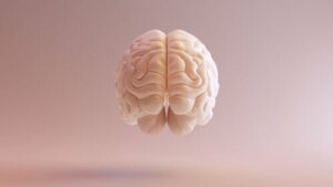
Neuroscience research has always been incredibly complex. Fortunately, some talented researchers take on the challenge so we can have a better understanding of the brain. One of those scientists was a German neurologist named Korbinian Brodmann, who divided the cerebral cortex into the Brodmann areas.
Brodmann dedicated his life to the study of anatomy and psychiatry. In 1901, he started to focus on neurobiology. During this period, he came up with the famous brain map that’s named after him.
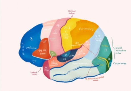
In the diagram of the Brodmann areas, you see how the different areas of the brain are divided according to their function and location. Keep reading to learn more about these complex and fascinating regions of the brain!
What are the Brodmann areas?
Brodmann made his map of the cerebral cortex around 1909. In it, he identified 52 different areas, and each one includes cytoarchitecture, blood flow, metabolism, and characteristic functions.
Brodmann’s goal was to create a topographical diagram of the cortex based on anatomical and functional characteristics. To do that, he made spatial divisions in the cortex. Through experimentation, these areas were correlated to different cortical functions.
Brodmann used the Nissl staining method during his neuroanatomy research to discover these areas. Interestingly, he didn’t do his research on exclusively human subjects. He also used monkeys.
Brodmann wasn’t the only scientist to divide the cerebral cortex into distinct areas. Constantin Von Economo and Georg N. Koskinas also did it and in much more detail. Nevertheless, Brodmann’s cortical map became the most widely used and is still a reference today.
While we understand now that there’s no exact division between the cortical areas and that there are interrelationships in the cerebral cortex, not independently functioning areas, this map is still very useful.
Motor areas
These are the regions that make up the motor cortex. This zone is in charge of processing information relating to muscle movement. That includes the generation, coordination, maintenance, and finalization of movements. The following zones are located in the motor cortex:
- The primary motor area (Brodmann area 4). This zone is characterized by a low arousal threshold. It’s responsible for sending signals to the muscles to start voluntary movements, usually simple ones.
- The supplementary motor area (Brodmann area 6). This area has a high arousal threshold. It’s responsible for involuntary postural movements. It also influences the organization of movement sequences of large muscle groups.
- The secondary motor area (Brodmann area 8). This is a premotor area and it works with area 6 to store movement patterns that come from past experiences. It’s also responsible for eye movement.
- The Broca Area (Brodmann 44 and 45). These zones are related to the necessary movements to produce language, i.e., gesticulation, intonation, and semantic processing. They play a crucial role in the production of speech and the written word.
Sensitive areas
These make up the somatosensorial region and they’re responsible for cerebral processing of sensorial phenomena (association and coordination of stimuli and compaction of prior stimuli with exterior stimuli).
Among other things, they process information from the tactile system and that related to body positioning. The following are sensitive regions:
- Primary somatosensory areas (Brodmann areas 1, 2, and 3). These are located between the parietal gyrus and the posterior part of the central parietal lobe. They’re responsible for touch and proprioception.
- Secondary somatosensory areas (Brodmann 5 and 7). 5 is responsible for tactile perception and 7 is a unifying area, related to sightless object recognition.
- Acoustic sensitivity areas. Area 41 detects changes in frequency and locates sound; area 42 participates in speech identification and recognition and processes information from the primary auditory cortex. Areas 20 and 21 recognize sounds and 22 perceives them.
- Taste sensitivity (Brodmann area 43). This area is on the posterior lip of the lateral sulcus and in the zone adjacent to the insula. Thanks to area 43, we can process information about flavor and taste.
- Vestibular sensitivity. Located in the posterior superior temporal gyrus and area 22. It plays a role in the perception of the positions of the body, movements of the head in space, and keeping your balance.
- Visual sensitivity. Brodmann area 17, located near the calcarine fissure and the occipital pole, processes visual content with a retinopathic distribution of visual representations. Areas 18 and 19 relate the information received from area 17 with past visual experiences to help recognize and appreciate what you can see.
Associative areas
These are multi-sensitive areas capable of making associations between different sensations and with motor-type areas. They relate to behavior, perceptive discrimination, and the interpretation of sensory experiences.
You can find them in three areas of the brain: the posterior parietal, temporal anterior, and prefrontal. Among them are:
- Prefrontal areas (Brodmann areas 9, 10, 11, and 12). These associate the experiences necessary to produce abstract ideas. They also have to do with executive functions, personality, and emotions.
- Angular area (Brodmann areas 39 and 40). These zones associate visual, proprioceptive, and tactile information to consolidate concepts of shape, size, and texture. They also play a role in the appreciation and awareness of images.
- Temporal anterior area. These areas store sensory experiences. When you stimulate this area of the brain, you remember objects or music you’ve previously experienced. Experts attribute the tapping of your foot along to the beat to area 38.
- Associative areas of language. Broca’s area is responsible for generating the movement necessary to produce speech. Wernicke’s area makes it possible to understand the written word and spoken language and associates sounds with concepts. The Exner center plays a role in written language, and the Dejerine and Luria areas are responsible for the appropriate organization of words.
Neuroscience research has always been incredibly complex. Fortunately, some talented researchers take on the challenge so we can have a better understanding of the brain. One of those scientists was a German neurologist named Korbinian Brodmann, who divided the cerebral cortex into the Brodmann areas.
Brodmann dedicated his life to the study of anatomy and psychiatry. In 1901, he started to focus on neurobiology. During this period, he came up with the famous brain map that’s named after him.
In the diagram of the Brodmann areas, you see how the different areas of the brain are divided according to their function and location. Keep reading to learn more about these complex and fascinating regions of the brain!
What are the Brodmann areas?
Brodmann made his map of the cerebral cortex around 1909. In it, he identified 52 different areas, and each one includes cytoarchitecture, blood flow, metabolism, and characteristic functions.
Brodmann’s goal was to create a topographical diagram of the cortex based on anatomical and functional characteristics. To do that, he made spatial divisions in the cortex. Through experimentation, these areas were correlated to different cortical functions.
Brodmann used the Nissl staining method during his neuroanatomy research to discover these areas. Interestingly, he didn’t do his research on exclusively human subjects. He also used monkeys.
Brodmann wasn’t the only scientist to divide the cerebral cortex into distinct areas. Constantin Von Economo and Georg N. Koskinas also did it and in much more detail. Nevertheless, Brodmann’s cortical map became the most widely used and is still a reference today.
While we understand now that there’s no exact division between the cortical areas and that there are interrelationships in the cerebral cortex, not independently functioning areas, this map is still very useful.
Motor areas
These are the regions that make up the motor cortex. This zone is in charge of processing information relating to muscle movement. That includes the generation, coordination, maintenance, and finalization of movements. The following zones are located in the motor cortex:
- The primary motor area (Brodmann area 4). This zone is characterized by a low arousal threshold. It’s responsible for sending signals to the muscles to start voluntary movements, usually simple ones.
- The supplementary motor area (Brodmann area 6). This area has a high arousal threshold. It’s responsible for involuntary postural movements. It also influences the organization of movement sequences of large muscle groups.
- The secondary motor area (Brodmann area 8). This is a premotor area and it works with area 6 to store movement patterns that come from past experiences. It’s also responsible for eye movement.
- The Broca Area (Brodmann 44 and 45). These zones are related to the necessary movements to produce language, i.e., gesticulation, intonation, and semantic processing. They play a crucial role in the production of speech and the written word.
Sensitive areas
These make up the somatosensorial region and they’re responsible for cerebral processing of sensorial phenomena (association and coordination of stimuli and compaction of prior stimuli with exterior stimuli).
Among other things, they process information from the tactile system and that related to body positioning. The following are sensitive regions:
- Primary somatosensory areas (Brodmann areas 1, 2, and 3). These are located between the parietal gyrus and the posterior part of the central parietal lobe. They’re responsible for touch and proprioception.
- Secondary somatosensory areas (Brodmann 5 and 7). 5 is responsible for tactile perception and 7 is a unifying area, related to sightless object recognition.
- Acoustic sensitivity areas. Area 41 detects changes in frequency and locates sound; area 42 participates in speech identification and recognition and processes information from the primary auditory cortex. Areas 20 and 21 recognize sounds and 22 perceives them.
- Taste sensitivity (Brodmann area 43). This area is on the posterior lip of the lateral sulcus and in the zone adjacent to the insula. Thanks to area 43, we can process information about flavor and taste.
- Vestibular sensitivity. Located in the posterior superior temporal gyrus and area 22. It plays a role in the perception of the positions of the body, movements of the head in space, and keeping your balance.
- Visual sensitivity. Brodmann area 17, located near the calcarine fissure and the occipital pole, processes visual content with a retinopathic distribution of visual representations. Areas 18 and 19 relate the information received from area 17 with past visual experiences to help recognize and appreciate what you can see.
Associative areas
These are multi-sensitive areas capable of making associations between different sensations and with motor-type areas. They relate to behavior, perceptive discrimination, and the interpretation of sensory experiences.
You can find them in three areas of the brain: the posterior parietal, temporal anterior, and prefrontal. Among them are:
- Prefrontal areas (Brodmann areas 9, 10, 11, and 12). These associate the experiences necessary to produce abstract ideas. They also have to do with executive functions, personality, and emotions.
- Angular area (Brodmann areas 39 and 40). These zones associate visual, proprioceptive, and tactile information to consolidate concepts of shape, size, and texture. They also play a role in the appreciation and awareness of images.
- Temporal anterior area. These areas store sensory experiences. When you stimulate this area of the brain, you remember objects or music you’ve previously experienced. Experts attribute the tapping of your foot along to the beat to area 38.
- Associative areas of language. Broca’s area is responsible for generating the movement necessary to produce speech. Wernicke’s area makes it possible to understand the written word and spoken language and associates sounds with concepts. The Exner center plays a role in written language, and the Dejerine and Luria areas are responsible for the appropriate organization of words.
All cited sources were thoroughly reviewed by our team to ensure their quality, reliability, currency, and validity. The bibliography of this article was considered reliable and of academic or scientific accuracy.
- Garey, L. J. (2006). Broadmann’s ‘Localisation in the Cerebral Cortex’. Springer.
This text is provided for informational purposes only and does not replace consultation with a professional. If in doubt, consult your specialist.