All About the Corpus Callosum
Written and verified by the psychologist Paula Villasante
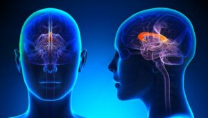
The corpus callosum, or callosal commissure, connects the left side of your brain to the right side. Each side is known as a hemisphere, and the connection allows information to pass between the two halves. Corpus callosum is Latin for “tough body.” It’s the largest connective pathway in the brain, as it’s made up of more than 200 million nerve fibers.
Very occasionally, a person will be born without a callosal commissure, also known as the agenesis of the corpus callosum. This causes many different physical and behavioral symptoms. Each side of the brain controls movement and feeling in the opposite half of the body, and the hemispheres also process information such as language.
Therefore, physical coordination and taking in complex information require both sides of the brain to work together. The callosal commissure acts as the connector. It sits in the center of the brain, measures approximately four inches in length, and looks exactly like the letter “C”.
The callosum usually forms in the brain between twelve and sixteen weeks after conception, near the end of the first trimester of pregnancy, developing throughout childhood. By the time a child is twelve years old, their corpus callosum will have finished developing.
It’ll then remain unchanged into adulthood and throughout the rest of their life. Until the 1950s, experts didn’t know the exact function of the callosal commissure. In 1955, Ronald Myers, a graduate student at the University of Chicago, proved its function was related to coordination and complex problem-solving.
What exactly does the corpus callosum do?
The callosal commissure is a large, C-shaped nerve fiber bundle found beneath the cerebral cortex. It stretches across the midline of the brain, connecting the left and right cerebral hemispheres.
Most importantly, it makes up the biggest collection of white matter tissue found in the brain. To understand the corpus callosum’s role, it’s vital to remember that the brain is divided into two cerebral hemispheres.
The hemispheres utterly differ from one another thanks to the medial longitudinal fissure. This fissure seems just like a long groove. On a large scale, the two hemispheres are nearly identical, though, on a microscopic and functional level, there are some differences. When they send information like sensory data to the brain, it typically receives it first in one hemisphere.
When you type on your keyboard, they send information about the feel of the keys, up from your right hand to the primary somatosensory cortex on the left side of your brain. Furthermore, they must then share the information with the right side of your brain as well.
That’s where the callosal commissure comes into play. It’s a large bundle of fibers that connects the left and right hemispheres, while perfectly carrying information from one cerebral hemisphere to the other.
Disorders
Did you know that some children are born without a callosal commissure? This leads to a rare disorder known as the agenesis of the corpus callosum. In fact, this disorder is estimated to affect around one in 3,000 people in the world.
The callosal commissure also damages. Disruptions to the development of the callosal commissure can occur between the fifth and sixteenth week of pregnancy. While there’s no certain cause, possible factors include:
- Prenatal infections or viruses, such as rubella.
- Genetic abnormalities, such as Andermann or Aicardi syndromes.
- Toxic metabolic conditions, such as fetal alcohol syndrome (heavy drinking or alcoholism during pregnancy).
- Something that prevents the callosal commissure from growing, such as a cyst in the brain.
The recessive genetic disorder can also cause callosal commissure problems. The parents can be carriers of the gene that causes the disorder, even if they don’t have the disorder themselves. Thus, a child with two carrier parents has a 25 percent chance of having problems with the callosal commissure. A child with two carrier parents has a 50 percent chance of becoming a carrier themselves.
This equally affects both males and females. However, thanks to an increase in scanning technology use, more children might receive a diagnosis in the future, such as magnetic resonance imaging (MRI). The callosal commissure’s presence isn’t essential for survival. Those who have problems with it will often fall behind their peers in development.
Children with agenesis may be blind, deaf, or never learn to walk or talk. Other children with agenesis can be very high-functioning. You often associate the term “high-functioning” with autism. When autistic people have strong verbal skills, and with a disproportionately high IQ level, they’re high-functioning.
It’s imperative to understand the disorder of the callosal commissure isn’t a disease or illness in itself. Not many people with agenesis of the corpus callosum lead healthy lives. However, it can also lead to medical problems. For example, seizures, which require medical intervention.
Conditions
A number of conditions are associated with the callosal commissure. Agenesis of the corpus callosum can sometimes cause other brain abnormalities, such as fluid. Are you ready to check out other conditions?
- Fluid in the skull, known as hydrocephalus.
- Arnold-Chiari malformation.
- Deep clefts in brain tissue.
- Failure of the forebrain to divide into lobes.
- Disorder of neural migration.
Likewise, agenesis of the corpus callosum also often happens in conjunction with spina bifida. Spina bifida is when the spine doesn’t fuse properly, leaving a defect in the spinal canal.
Symptoms and diagnosis
Did you know that experts diagnose agenesis of the corpus callosum in the first two years of a person’s life? An epileptic seizure is often the first sign of brain dysfunction. In mild cases, the disorder can go undetected for some years. Experts require a brain scan to confirm problems with the callosal commissure. The brain tests can include the following:
- Prenatal ultrasound.
- Computerized tomography (CT scan).
- MRI scan.
Research on callosal disorders is ongoing, and there are some common characteristics of agenesis, which vary from person to person. As a matter of fact, it can be broken down into four categories, such as physical, cognitive, developmental, and social.
Physical characteristics include low muscle tone and cognitive characteristics include a lack of ability in assessing risk. Developmental characteristics include poor coordination and social characteristics include both immaturity and hyperactivity.
Fun facts
Do you want to understand the corpus callosum better? Check out these few fun facts to help you remember:
- The callosum is part of the brain in humans and other eutherian mammals.
- It’s the biggest connective pathway in the brain.
- Over 200 million nerve fibers connect the left and right hemispheres.
- Until around 1950, its function was a complete mystery.
- On rare occasions, it’s completely absent at birth.
- Experts cut the callosum surgically in order to treat epilepsy.
Split-brain patients
In the second half of the 20th century, Roger Sperry, Gazzaniga, and others studied patients with a major issue. Doctors severed their callosal commissure in the surgery corpus callosotomy.
They normally undertake the procedure as a last-resort treatment of epilepsy, and it can stop seizures from spreading from one brain hemisphere to another.
The world now knows groundbreaking patients as split-brain patients. Surprisingly, the doctors can complete a corpus callosotomy without severe side effects, though the side effects that do appear are often related to language.
Sperry and Gazzaniga explored language deficits in callosotomy patients in detail. In the process, they learned some interesting things, such as how language centers are distributed across the cerebral hemispheres.
They also learned how the callosal commissure facilitates communication between the two sides of the brain. Sperry and Gazzaniga presented split-brain patients with visual stimuli, though only to one eye at a time.
For example, they presented an image of a flower to the right eye and covered the left eye. Their split-brain patients’ research amazed researchers. Patients presented with a visual image to only their left eye, couldn’t name the object in the image.
Hypothesis and results
Sperry and colleagues devised a brilliant hypothesis. They believed it occurred because information for the visual field travels to the opposite side of the brain. If they show the object to the left eye, information travels to the brain’s right side.
Normally, they would then share this information with the opposite hemisphere via the corpus callosum. Recent research also suggests that split-brain people couldn’t name the object either.
If they only showed it to the left eye, the visual information wasn’t reaching the left side of the brain. The left side of our brain is where the important language centers locate. Much of what we know about one cerebral hemisphere dominating isn’t true.
Experts probably exaggerate its dominance in managing a capacity or particular skill. A creative person doesn’t have an overall bias toward thinking with the right side of her brain. On the other hand, their skills are spread evenly throughout both hemispheres. Language seems to be an exception.
In most people, speech comes from the left hemisphere. They consider the left hemisphere to be the dominant one for language. According to Sperry and Gazzaniga, language centers are on the left side of the brain.
When they present an image to the left eye of a split-brain patient, the patient’s language areas aren’t privy to the visual information. The information travels to the right hemisphere, though doesn’t cross back over to the left thanks to the severed callosal commissure. Hence, they limit the ability to place a name on the object.
Outlook
The studies proved the importance of the left hemisphere in language processing. It also proved their relevance in bridging the two cerebral hemispheres. Furthermore, they also demonstrated the brain’s versatility and resilience. In most split-brain patients other tracts like the anterior commissure help.
It still carries enough information between the cerebral hemispheres, allowing functionality to be somewhat normal. If the callosal commissure didn’t grow properly during fetal development, it never will, and there’s no direct treatment for agenesis. Doctors will identify and potentially treat symptoms as they occur.
In short, there’s treatment available for many medical conditions associate with agenesis of the corpus callosum. Therapy and counseling can also help to perfectly improve language and social skills. A skilled doctor can help you outline all of the treatment options available in individual cases.
All cited sources were thoroughly reviewed by our team to ensure their quality, reliability, currency, and validity. The bibliography of this article was considered reliable and of academic or scientific accuracy.
- Función del cuerpo calloso: Implicaciones optométricas. Retrieved from http://www.acotv.org/es/blog/174-funcion-del-cuerpo-calloso-implicaciones-optometricas.html
-
Kwinta, P., Herman-Sucharska, I., Leśniak, A., Klimek, M., Karcz, P., Durlak, W., … & Pietrzyk, J. J. (2015). Relationship between stereoscopic vision, visual perception, and microstructure changes of corpus callosum and occipital white matter in the 4-year-old very low birth weight children. BioMed research international, 2015.
-
Renard, D., Castelnovo, G., Campello, C., Bouly, S., Le Floch, A., Thouvenot, E., … & Taieb, G. (2014). An MRI review of acquired corpus callosum lesions. J Neurol Neurosurg Psychiatry, 85(9), 1041-1048.
This text is provided for informational purposes only and does not replace consultation with a professional. If in doubt, consult your specialist.