Characteristics of the Cerebral Ventricular System
The nervous system is sort of like an orchestra conductor. Its various parts interact with one another and are responsible for various functions. Thus, some of its systems ensure its proper functioning. This is the case of the cerebral ventricular system which basically consists of small reservoirs that are connected to each other. It’s kind of like a sewage system.
The encephalous is inside the brain. Furthermore, the ventricular system, composed of four ventricles, is inside the brain. This system is in charge of maintaining, protecting, and structuring the brain. Although it’s quite important, few have heard of it.
This article will take you on a tour of the ventricular system and define what it consists of and explain the difference between each of its cavities and their functions. It’ll also explore some of its related alterations.
“Humor is by far the most significant activity of the human brain.”
-Edward de Bono-
What’s the ventricular system and what’s its origin?
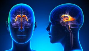
The set of cerebral ventricles is the ventricular system, composed of structures and interconnected cavities. The cerebrospinal fluid (CSF) that originates in these cavities circulates and keeps the brain and spinal cord moist.
The ventricular system develops parallel to the rest of the central nervous system, facilitating the circulation of CSF during the process. The differentiation of the optic ventricle begins approximately on the 26th day of embryonic development. Later, an evagination begins in the midline of the midbrain, which later forms the cerebral aqueduct.
The development of the interventricular foramen begins around the sixth week and initiates the formation of the choroid plexus of the lateral ventricles. Then, the grooves and segmentation become more noticeable.
Later, there’s a growth of the medial and lateral ventricular eminence and the spherical shape of the lateral ventricle becomes similar to a “c”. Thus, the horns of the lateral ventricles become more pronounced. In addition, a small sac forms on the diencephalic floor, which will be the third ventricle. The choroidal sacs of the fourth ventricle advance in their formation in the meantime.
Finally, during the seventh and eighth week, the horns finish defining themselves and constitute the definitive vesicular form. The isthmic part compresses due to the growing cerebellum and numerous villi extend into the midline.
Structure
The ventricular system has four ventricles interconnected through channels and openings. Here’s a description of each of its parts:
- The lateral ventricles (first and second ventricle) are in both cerebral hemispheres and have an anterior pole oriented to the frontal lobe and a posterior pole directed towards the temporal lobe. Also, they’re connected through the third ventricle by the Monro ventricular orifice. They’re C-shaped and their volume increases throughout life.
- The third ventricle consists of a thin, flattened cavity located between the thalamus and crossed by the interthalamic commissure. Its connection with the rest of the system is through the cerebral aqueduct. It has two protrusions:
- The supra-optic recess, located above the optic chiasm.
- The infundibular recess, located above the optic stem.
- In addition, the fourth ventricle extends from the mesencephalic aqueduct to the central channel at the top of the spinal cord. Its floor is comprised of the rhomboidal fossa and communicates with the central channel through Luschka and Magendie holes, from which the CSF comes out into the subarachnoid space. In addition, it connects to the subarachnoid cisterns that allow the CSF to reach the subarachnoid space.
The ventricles continue through the ependymal canal upon reaching the spinal cord. This cavity begins at the end of the fourth ventricle and runs internally through the cord until it reaches the first vertebra in the lumbar area.
Functions of the cerebral ventricular system
The ventricular system performs different functions:
- CSF production. This is its main function, although it shares it with other structures, such as the subarachnoid space.
- Preservation of the brain. The CSF helps maintain the brain’s internal homeostasis, maintaining adequate intracranial pressure. In addition, this liquid helps eliminate waste and this allows the brain to maintain an adequate environment for its functioning.
- Brain buoyancy. The brain floats thanks to the CSF and this contributes to the reduction of its weight from about 49 to one ounce.
- Defense. The CSF defends the brain against external agents that could be dangerous. It also increases its degree of protection against trauma.
Ventricular system alterations
The ventricular system can suffer alterations of various types. Here are some of them:
- Hydrocephalus. This is a disorder in which there’s an overproduction of CSF. It causes high intracranial pressure as it evolves and can lead to brain atrophy, metabolic and cognitive disorders, and even death.
- Meningitis. It consists of the inflammation of the meninges due to viral, fungal, or bacterial infection. This inflammation produces an increase in intracranial pressure, making the circulation of the CSF difficult and leading to symptoms such as headaches, nausea, sensitivity to light, fever, and cognitive impairment, among others.
- Ventriculitis. This is the inflammation of the cerebral ventricles that raises the intracranial pressure and alters the normal circulation of the CSF and the correct functioning of the vascular system. It can present with hydrocephalus and produce encephalitis or inflammation of the brain, among other pathologies.
- Alzheimer’s disease. The deterioration and death of neurons increases as this condition progresses and causes a decrease in neuronal density. The space vacated by the subsequent decrease in volume is occupied by the ventricles, which progressively expand.
- Schizophrenia. Some studies suggest that people with this condition have larger ventricles in the brain. In fact, Jordi E. Obiols and Marta Carulla from the Autonomous University of Barcelona, published an article in the journal Behavior Psychology. In it, they suggest that schizophrenic patients present ventricular dilatation and cortical shrinkage. Thus, this disease may start from neurodevelopment.

In conclusion
As you can see, the cerebral ventricular system is fundamental, and its malfunctioning causes serious alterations in the body.
As we mentioned at the beginning, the cerebral ventricular system is a kind of shield that protects you from adversity. Furthermore, it does everything possible to maintain internal balance and rejects harmful agents.
The nervous system is sort of like an orchestra conductor. Its various parts interact with one another and are responsible for various functions. Thus, some of its systems ensure its proper functioning. This is the case of the cerebral ventricular system which basically consists of small reservoirs that are connected to each other. It’s kind of like a sewage system.
The encephalous is inside the brain. Furthermore, the ventricular system, composed of four ventricles, is inside the brain. This system is in charge of maintaining, protecting, and structuring the brain. Although it’s quite important, few have heard of it.
This article will take you on a tour of the ventricular system and define what it consists of and explain the difference between each of its cavities and their functions. It’ll also explore some of its related alterations.
“Humor is by far the most significant activity of the human brain.”
-Edward de Bono-
What’s the ventricular system and what’s its origin?
The set of cerebral ventricles is the ventricular system, composed of structures and interconnected cavities. The cerebrospinal fluid (CSF) that originates in these cavities circulates and keeps the brain and spinal cord moist.
The ventricular system develops parallel to the rest of the central nervous system, facilitating the circulation of CSF during the process. The differentiation of the optic ventricle begins approximately on the 26th day of embryonic development. Later, an evagination begins in the midline of the midbrain, which later forms the cerebral aqueduct.
The development of the interventricular foramen begins around the sixth week and initiates the formation of the choroid plexus of the lateral ventricles. Then, the grooves and segmentation become more noticeable.
Later, there’s a growth of the medial and lateral ventricular eminence and the spherical shape of the lateral ventricle becomes similar to a “c”. Thus, the horns of the lateral ventricles become more pronounced. In addition, a small sac forms on the diencephalic floor, which will be the third ventricle. The choroidal sacs of the fourth ventricle advance in their formation in the meantime.
Finally, during the seventh and eighth week, the horns finish defining themselves and constitute the definitive vesicular form. The isthmic part compresses due to the growing cerebellum and numerous villi extend into the midline.
Structure
The ventricular system has four ventricles interconnected through channels and openings. Here’s a description of each of its parts:
- The lateral ventricles (first and second ventricle) are in both cerebral hemispheres and have an anterior pole oriented to the frontal lobe and a posterior pole directed towards the temporal lobe. Also, they’re connected through the third ventricle by the Monro ventricular orifice. They’re C-shaped and their volume increases throughout life.
- The third ventricle consists of a thin, flattened cavity located between the thalamus and crossed by the interthalamic commissure. Its connection with the rest of the system is through the cerebral aqueduct. It has two protrusions:
- The supra-optic recess, located above the optic chiasm.
- The infundibular recess, located above the optic stem.
- In addition, the fourth ventricle extends from the mesencephalic aqueduct to the central channel at the top of the spinal cord. Its floor is comprised of the rhomboidal fossa and communicates with the central channel through Luschka and Magendie holes, from which the CSF comes out into the subarachnoid space. In addition, it connects to the subarachnoid cisterns that allow the CSF to reach the subarachnoid space.
The ventricles continue through the ependymal canal upon reaching the spinal cord. This cavity begins at the end of the fourth ventricle and runs internally through the cord until it reaches the first vertebra in the lumbar area.
Functions of the cerebral ventricular system
The ventricular system performs different functions:
- CSF production. This is its main function, although it shares it with other structures, such as the subarachnoid space.
- Preservation of the brain. The CSF helps maintain the brain’s internal homeostasis, maintaining adequate intracranial pressure. In addition, this liquid helps eliminate waste and this allows the brain to maintain an adequate environment for its functioning.
- Brain buoyancy. The brain floats thanks to the CSF and this contributes to the reduction of its weight from about 49 to one ounce.
- Defense. The CSF defends the brain against external agents that could be dangerous. It also increases its degree of protection against trauma.
Ventricular system alterations
The ventricular system can suffer alterations of various types. Here are some of them:
- Hydrocephalus. This is a disorder in which there’s an overproduction of CSF. It causes high intracranial pressure as it evolves and can lead to brain atrophy, metabolic and cognitive disorders, and even death.
- Meningitis. It consists of the inflammation of the meninges due to viral, fungal, or bacterial infection. This inflammation produces an increase in intracranial pressure, making the circulation of the CSF difficult and leading to symptoms such as headaches, nausea, sensitivity to light, fever, and cognitive impairment, among others.
- Ventriculitis. This is the inflammation of the cerebral ventricles that raises the intracranial pressure and alters the normal circulation of the CSF and the correct functioning of the vascular system. It can present with hydrocephalus and produce encephalitis or inflammation of the brain, among other pathologies.
- Alzheimer’s disease. The deterioration and death of neurons increases as this condition progresses and causes a decrease in neuronal density. The space vacated by the subsequent decrease in volume is occupied by the ventricles, which progressively expand.
- Schizophrenia. Some studies suggest that people with this condition have larger ventricles in the brain. In fact, Jordi E. Obiols and Marta Carulla from the Autonomous University of Barcelona, published an article in the journal Behavior Psychology. In it, they suggest that schizophrenic patients present ventricular dilatation and cortical shrinkage. Thus, this disease may start from neurodevelopment.
In conclusion
As you can see, the cerebral ventricular system is fundamental, and its malfunctioning causes serious alterations in the body.
As we mentioned at the beginning, the cerebral ventricular system is a kind of shield that protects you from adversity. Furthermore, it does everything possible to maintain internal balance and rejects harmful agents.
All cited sources were thoroughly reviewed by our team to ensure their quality, reliability, currency, and validity. The bibliography of this article was considered reliable and of academic or scientific accuracy.
- Bear, M. F. Connors, B. W., Paradiso, M.A., Nuin, X. U., Guillén, X. V. & Sol Jaquotot, M. J. (2008). Neurociencias: la exploración del cerebro. Wolters Kluwer/Lippincott Williams & Wikins.
- Obiols, J.E., & Carulla, M. (1998). Bases biològicas de la esquizofrenia: Aspectos neuroquímicos y neuroanatómicos. Psicología Conductual, 6(1), 5-27.
This text is provided for informational purposes only and does not replace consultation with a professional. If in doubt, consult your specialist.