The Spinal Cord: Anatomy and Physiology
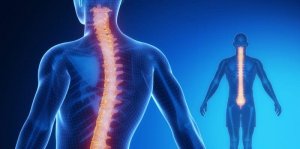
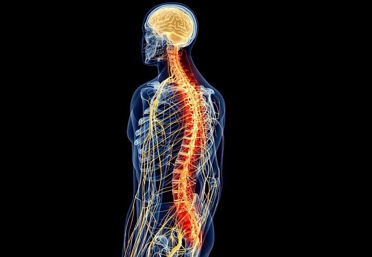
The spinal cord is part of the Central Nervous System (CNS) along with the encephalon. Its extension goes from the occipital foramen of the skull to approximately the first lumbar vertebra.
31 spinal nerves are connected along the spinal cord. It is composed of a core of gray matter where the neuronal bodies are located. This, in turn, is surrounded by white matter where the axons are located. Interestingly, the distribution of gray and white matter in the spinal cord is the opposite of that of the brain. On the other hand, vertebrae, support ligaments,meninges, and cerebrospinal fluid protect the spinal cord.
The spinal cord performs various functions. It is responsible for receiving and processing (in a superficial way) information from the senses. It also sends motor information from the brain. These functions are of vital importance. An injury can cause serious effects such as paralysis at motor level or loss of sensitivity.
Gray substance
This gray matter, unlike that of the brain, is located in the inner part of the spinal cord. It is the place where neuronal bodies are located and where information is processed. It is configured in different antlers: ventral, dorsal, lateral, and intermediate.
- Dorsal horn: responsible for information from the senses.
- Intermediate zone: place where the inter-neurons that link neurons are found. They are association neurons.
- Lateral horn: only found at the thoracic and lumbar level. It is responsible for the homeostasis of the body, regulating the automatic nervous system.
- Ventral horn: is responsible for motor information.
In addition, inside this gray substance there are several nuclei with different functions:
- I-IV: responsible for the exteroceptive sensations. They record the sensations they receive from external stimuli, such as light.
- V-VI: responsible for proprioceptive sensations. They report stimuli generated internally.
- VIII: carries out the relay between mesencephalon and cerebellum. It is the place where the neurons coming from the mesencephalon take over to go to the cerebellum and vice versa.
- IX: main motor area. It is the place where the descending neuronal bodies that come from the motor cortex and direct impulses of movement.
- X: nucleus that surrounds the central duct and contains neuroglia, which are support neurons.
The gray matter of the spinal cord is primarily a relay site for motor and sensory information. It also has to make a quick judgement on the information before it even reaches its destination. The latter is necessary to activate the reflexes mechanism in emergency situations, such as receiving a very painful stimulus.
White substance
The white matter of the spinal cord is the place where the fibers (axons) exist that send information up and down. Its main function is to send information. Like the substantia nigra, it is also divided into different parts. In this case, columns:
- Dorsal column: sends somatic information.
- Ventral and lateral column: the efferent pathways that are responsible for sending information from the brain to the muscles. They are part of the motor system.
Within the white substance there are different routes – ascending and descending. The tracts take the name of the two structures between which the information circulates. Furthermore, each of the tracts sends different information.
- Gracile and Cuneate: deal with the discriminative touch and movements of the hands.
- Anterior and posterior spinocerebellar: unconscious movements coming from muscles, skin joints, and subcutaneous tissue.
- Spino-olivar: although this tract has been located, its function is not exactly known.
- Spinothalamic lateral: painful and thermal sensations.
- Spinotectal: afferent information for spinovisual reflexes.
- Anterior spinothalamic: light touch and pressure.
- Anterior and lateral corticospinal: confers agility and rapidity to movements.
- Tectospinal: participates in movements from visual stimuli.
- Vestibulospinal: responsible for maintaining balance.
- Olivospinal: influences the activity of motor neurons.
- Rubrospinal: inhibits the activity of the extensor muscles.
Thus, the white matter of the spinal cord is responsible for the transmission of motor and sensory information in a wide range of movements and sensations, communicating between multiple areas.
Ascending (sensory)
The ascending pathways, as the name indicates, are responsible for sending the information collected by the external senses (exteroceptive information) or internal stimuli (proprioceptive) to the cerebral cortex where deeper processing will be done. Most of the ascending pathways end in the thalamus, with the exception of the olfactory stimuli that go directly to the olfactory bulb.

They are ascending, centripetal, born from the periphery, and provide information to higher centers. Some of the nerve fibers serve to link different segments of the spinal cord. Meanwhile, others ascend from the cord to the upper centers and thus connect the spinal cord to the brain. They conduct information that may or may not reach the conscience.
In its simplest form, the pathway that ascends to consciousness consists of three neurons. However, some afferent pathways use more or less. Many of the neurons present in the ascending pathways branch out and others participate in reflex muscle activity.
They are those routes that direct information from somatic receptors. There are two main ways this information travels:
- Nocioceptive pathway: conducts pain and temperature information.
- Mechanical path: conveys information about superficial and deep touch, proprioception, and vibration.
Descending (motor)
The pyramidal pathways are those descending (motor) nerve pathways that pass through the pyramids. They are in charge of voluntary, fast, agile, fine, and precise movement. There are three neurons involved in sending information to execute a movement. They follow this circuit:
- Neuron 1: neuron located in the pre-central and pre-motor cortex.
- Neuron 2: does not always exist in the path. It is an interneuron or internuncial neuron.
- Neuron 3: located on the anterior horn of the spinal cord. All the pyramidal tracts end up presenting contralaterality, which means that a lesion in the right motor cortex will cause an injury in the left part of the body, for example.
The extrapyramidal route is responsible for involuntary movements and comes from some subcortical structure, travelling to the spinal cord. It regulates the execution of involuntary movements (walking, posture, muscle tone, alertness level, and instinctive behavior). Unlike the pyramidal system, it does not start in the cerebral cortex, but rather in various subcortical structures.
Another critical function of the descending motor pathways is to modulate the reflex circuits in the spinal cord. The adaptability of the spinal reflexes can changed depending on the behavioral context. Sometimes the gain (strength) or even the sign (extension vs. flexion) must be changed so that the movement adapts to the circumstances. Descending pathways are responsible for controlling these variables.
Reflexes of the spinal cord
There are movements that we make unconsciously before the sensory information of the stimulus that causes the movement reaches the brain. These are reflex movements such as removing the hand from a painful source or closing the eyes when we hear a loud noise. We do not control these movements.
The reflex is the simplest circuit of the nervous system. It starts in the receptors, which are structures that transform the energy of the stimulus to an electrical change in the peripheral afferent nerves. These drive the impulses to the integrating center, the interneuron. Then, the information passes to the efferent motor neurons, so that the effector (muscle) performs the reflex movement.
These movements are thanks to the reflex arc. The soma of the neuron is located in the ganglion of the posterior root, and passes through the dorsal where it communicates with the interneuron, which is the association neuron that integrates the information and passes it to the motor neuron in the ventral horn to leave the ventral root and direct the nervous impulse to the muscle for its contraction.
Bibliography
Carlson R. (2014). Fisiología de la conducta. Madrid, España: Pearson educación
Tortora G. J., Derrickson B.(2013). Principios de anatomía y fisiología. Buenos Aires: Editorial Médica Panamericana
Kandell E.R., Schwartz J.H. y Jessell T.M.(2001). Principios de Neurociencia. Madrid: McGraw-Hill/Interamericana.
The spinal cord is part of the Central Nervous System (CNS) along with the encephalon. Its extension goes from the occipital foramen of the skull to approximately the first lumbar vertebra.
31 spinal nerves are connected along the spinal cord. It is composed of a core of gray matter where the neuronal bodies are located. This, in turn, is surrounded by white matter where the axons are located. Interestingly, the distribution of gray and white matter in the spinal cord is the opposite of that of the brain. On the other hand, vertebrae, support ligaments,meninges, and cerebrospinal fluid protect the spinal cord.
The spinal cord performs various functions. It is responsible for receiving and processing (in a superficial way) information from the senses. It also sends motor information from the brain. These functions are of vital importance. An injury can cause serious effects such as paralysis at motor level or loss of sensitivity.
Gray substance
This gray matter, unlike that of the brain, is located in the inner part of the spinal cord. It is the place where neuronal bodies are located and where information is processed. It is configured in different antlers: ventral, dorsal, lateral, and intermediate.
- Dorsal horn: responsible for information from the senses.
- Intermediate zone: place where the inter-neurons that link neurons are found. They are association neurons.
- Lateral horn: only found at the thoracic and lumbar level. It is responsible for the homeostasis of the body, regulating the automatic nervous system.
- Ventral horn: is responsible for motor information.
In addition, inside this gray substance there are several nuclei with different functions:
- I-IV: responsible for the exteroceptive sensations. They record the sensations they receive from external stimuli, such as light.
- V-VI: responsible for proprioceptive sensations. They report stimuli generated internally.
- VIII: carries out the relay between mesencephalon and cerebellum. It is the place where the neurons coming from the mesencephalon take over to go to the cerebellum and vice versa.
- IX: main motor area. It is the place where the descending neuronal bodies that come from the motor cortex and direct impulses of movement.
- X: nucleus that surrounds the central duct and contains neuroglia, which are support neurons.
The gray matter of the spinal cord is primarily a relay site for motor and sensory information. It also has to make a quick judgement on the information before it even reaches its destination. The latter is necessary to activate the reflexes mechanism in emergency situations, such as receiving a very painful stimulus.
White substance
The white matter of the spinal cord is the place where the fibers (axons) exist that send information up and down. Its main function is to send information. Like the substantia nigra, it is also divided into different parts. In this case, columns:
- Dorsal column: sends somatic information.
- Ventral and lateral column: the efferent pathways that are responsible for sending information from the brain to the muscles. They are part of the motor system.
Within the white substance there are different routes – ascending and descending. The tracts take the name of the two structures between which the information circulates. Furthermore, each of the tracts sends different information.
- Gracile and Cuneate: deal with the discriminative touch and movements of the hands.
- Anterior and posterior spinocerebellar: unconscious movements coming from muscles, skin joints, and subcutaneous tissue.
- Spino-olivar: although this tract has been located, its function is not exactly known.
- Spinothalamic lateral: painful and thermal sensations.
- Spinotectal: afferent information for spinovisual reflexes.
- Anterior spinothalamic: light touch and pressure.
- Anterior and lateral corticospinal: confers agility and rapidity to movements.
- Tectospinal: participates in movements from visual stimuli.
- Vestibulospinal: responsible for maintaining balance.
- Olivospinal: influences the activity of motor neurons.
- Rubrospinal: inhibits the activity of the extensor muscles.
Thus, the white matter of the spinal cord is responsible for the transmission of motor and sensory information in a wide range of movements and sensations, communicating between multiple areas.
Ascending (sensory)
The ascending pathways, as the name indicates, are responsible for sending the information collected by the external senses (exteroceptive information) or internal stimuli (proprioceptive) to the cerebral cortex where deeper processing will be done. Most of the ascending pathways end in the thalamus, with the exception of the olfactory stimuli that go directly to the olfactory bulb.
They are ascending, centripetal, born from the periphery, and provide information to higher centers. Some of the nerve fibers serve to link different segments of the spinal cord. Meanwhile, others ascend from the cord to the upper centers and thus connect the spinal cord to the brain. They conduct information that may or may not reach the conscience.
In its simplest form, the pathway that ascends to consciousness consists of three neurons. However, some afferent pathways use more or less. Many of the neurons present in the ascending pathways branch out and others participate in reflex muscle activity.
They are those routes that direct information from somatic receptors. There are two main ways this information travels:
- Nocioceptive pathway: conducts pain and temperature information.
- Mechanical path: conveys information about superficial and deep touch, proprioception, and vibration.
Descending (motor)
The pyramidal pathways are those descending (motor) nerve pathways that pass through the pyramids. They are in charge of voluntary, fast, agile, fine, and precise movement. There are three neurons involved in sending information to execute a movement. They follow this circuit:
- Neuron 1: neuron located in the pre-central and pre-motor cortex.
- Neuron 2: does not always exist in the path. It is an interneuron or internuncial neuron.
- Neuron 3: located on the anterior horn of the spinal cord. All the pyramidal tracts end up presenting contralaterality, which means that a lesion in the right motor cortex will cause an injury in the left part of the body, for example.
The extrapyramidal route is responsible for involuntary movements and comes from some subcortical structure, travelling to the spinal cord. It regulates the execution of involuntary movements (walking, posture, muscle tone, alertness level, and instinctive behavior). Unlike the pyramidal system, it does not start in the cerebral cortex, but rather in various subcortical structures.
Another critical function of the descending motor pathways is to modulate the reflex circuits in the spinal cord. The adaptability of the spinal reflexes can changed depending on the behavioral context. Sometimes the gain (strength) or even the sign (extension vs. flexion) must be changed so that the movement adapts to the circumstances. Descending pathways are responsible for controlling these variables.
Reflexes of the spinal cord
There are movements that we make unconsciously before the sensory information of the stimulus that causes the movement reaches the brain. These are reflex movements such as removing the hand from a painful source or closing the eyes when we hear a loud noise. We do not control these movements.
The reflex is the simplest circuit of the nervous system. It starts in the receptors, which are structures that transform the energy of the stimulus to an electrical change in the peripheral afferent nerves. These drive the impulses to the integrating center, the interneuron. Then, the information passes to the efferent motor neurons, so that the effector (muscle) performs the reflex movement.
These movements are thanks to the reflex arc. The soma of the neuron is located in the ganglion of the posterior root, and passes through the dorsal where it communicates with the interneuron, which is the association neuron that integrates the information and passes it to the motor neuron in the ventral horn to leave the ventral root and direct the nervous impulse to the muscle for its contraction.
Bibliography
Carlson R. (2014). Fisiología de la conducta. Madrid, España: Pearson educación
Tortora G. J., Derrickson B.(2013). Principios de anatomía y fisiología. Buenos Aires: Editorial Médica Panamericana
Kandell E.R., Schwartz J.H. y Jessell T.M.(2001). Principios de Neurociencia. Madrid: McGraw-Hill/Interamericana.
This text is provided for informational purposes only and does not replace consultation with a professional. If in doubt, consult your specialist.