Occipital Lobe: Structure and Functions

Written and verified by the psychologist Valeria Sabater
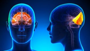
Take a deep breath and look around you right now. Don’t rush it. The world is full of beauty; of small nuances that make up our exciting reality. The reason why most of us are able to perceive the visual stimuli that surround us in full detail is, basically, the occipital lobe. It’s the area of our brain located at the nape of our neck.
It’s interesting how this region, the smallest of the four cerebral lobes, is the one that makes the most difference in your daily life. Its main purpose may seem simple at first: to receive information through your eyes to later process it and send it to the frontal lobe so it emits a response.
Now, if you carefully analyze that first look you took at the beginning of this article, you’ll realize it’s anything but easy. When your brain receives a stimulus, it performs many processes. It analyzes distances with regard to your position, movements, sizes, and also processes light (color).
All of this is something you do unconsciously. It entails a high level of neurological sophistication. Absolute precision where the occipital lobe allows you to move effectively in your daily routine. It’s small but highly specialized and effective. Let’s find out more about it.
“The brain is the most complicated organ in the universe. We’ve learned a lot about other human organs. We know how the heart pumps and how the kidney does what it does. To a certain degree, we’ve read the letters of the human genome. But the brain has 100 billion neurons. Each one of those has about 10,000 connections.”
-Francis Collins-
Occipital Lobe: Location and Structure
The occipital lobe is located in the back of the cerebral cortex. It occupies about 12% of the neocortex and is, in turn, connected to the primary visual cortex and of association and with the calcarine sulcus, a gyrus that’s just inside it. All these connections elevate it to a neural center of human vision and visual perception.
Note that, as your four cerebral lobes, it has a left and right hemisphere. However, each one of them is isolated from the other by the separation of the longitudinal fissure, relying in turn on the cerebellum and dura mater.
Functions and Areas of the Occipital Lobe

Your understanding of the world is based almost exclusively on your visual perception. Moreover, the occipital lobe is constantly processing visual stimuli, analyzing distances, shapes, colors, and movements.
Everything that goes through the retina passes through this analysis and processing center, then the information goes straight to the cerebral cortex. However, in order to execute this transfer of information, it must first go through the following areas:
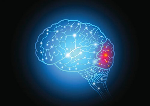
- Brodmann area 17 or Primary visual cortex (V1). Located in the rearmost region of the occipital lobe. In the event of an injury in this region, a person would be unable to see because they couldn’t process any stimulus, even if their eyes and retinas were in perfect condition.
- Brodmann area 18 or Secondary visual cortex (V2). Here, the prestriate cortex and the inferior temporal cortex extend. The first, in addition to receiving information from the primary visual area, is also responsible for stimulating memory. This allows you to associate visual stimuli with other stimuli you’ve previously seen. The inferior temporal cortex helps you recognize what you see.
- Brodmann area 19 or Associative visual cortex (V3, V4, and V5). This area receives information from the previous structures. Its main function is to process color and movement.
Occipital Lobe Injuries
Falls, traffic accidents, strokes, infections… The many conditions that may lead to an injury or an alteration in the occipital lobe can be severe and even permanent, as revealed by a study carried out at Nihon University in Tokyo, Japan.
These are the most common consequences of an occipital lobe injury:
Cortical Blindness
Cerebral visual impairment or cortical blindness is a consequence of a bilateral lesion in the primary visual cortex. In addition, people with this problem can only see diffused shapes. Vague stimuli of which they can’t recognize form or color, be it a particular situation or if something moves (or not).
Visual Hallucinations
An injury to this area of your brain can also lead to visual hallucinations. An injured person may perceive everything around them, but in a twisted manner, with strange colors and distorted sizes.
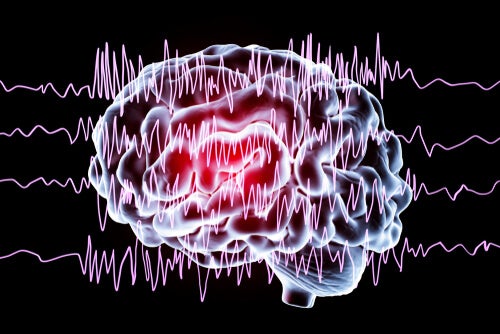
Epilepsy
The Department of Neurology at Yale School of Medicine explained the relationship between the occipital lobe and epilepsy through a study. A person may experience an epileptic seizure by overstimulating the neurons in this area as a result of being exposed to an intense flash of light.
This is, therefore, another type of epilepsy, one related to this particular area of our brain.
To conclude, note that the occipital lobe may be related to other processes beyond vision.
Furthermore, neurologists suspect it could also have something to do with memory. However, to this day, there are no conclusive studies. Perhaps in the coming years, and as researchers uncover more mysteries of the human brain, there’ll be more answers.
Take a deep breath and look around you right now. Don’t rush it. The world is full of beauty; of small nuances that make up our exciting reality. The reason why most of us are able to perceive the visual stimuli that surround us in full detail is, basically, the occipital lobe. It’s the area of our brain located at the nape of our neck.
It’s interesting how this region, the smallest of the four cerebral lobes, is the one that makes the most difference in your daily life. Its main purpose may seem simple at first: to receive information through your eyes to later process it and send it to the frontal lobe so it emits a response.
Now, if you carefully analyze that first look you took at the beginning of this article, you’ll realize it’s anything but easy. When your brain receives a stimulus, it performs many processes. It analyzes distances with regard to your position, movements, sizes, and also processes light (color).
All of this is something you do unconsciously. It entails a high level of neurological sophistication. Absolute precision where the occipital lobe allows you to move effectively in your daily routine. It’s small but highly specialized and effective. Let’s find out more about it.
“The brain is the most complicated organ in the universe. We’ve learned a lot about other human organs. We know how the heart pumps and how the kidney does what it does. To a certain degree, we’ve read the letters of the human genome. But the brain has 100 billion neurons. Each one of those has about 10,000 connections.”
-Francis Collins-
Occipital Lobe: Location and Structure
The occipital lobe is located in the back of the cerebral cortex. It occupies about 12% of the neocortex and is, in turn, connected to the primary visual cortex and of association and with the calcarine sulcus, a gyrus that’s just inside it. All these connections elevate it to a neural center of human vision and visual perception.
Note that, as your four cerebral lobes, it has a left and right hemisphere. However, each one of them is isolated from the other by the separation of the longitudinal fissure, relying in turn on the cerebellum and dura mater.
Functions and Areas of the Occipital Lobe
Your understanding of the world is based almost exclusively on your visual perception. Moreover, the occipital lobe is constantly processing visual stimuli, analyzing distances, shapes, colors, and movements.
Everything that goes through the retina passes through this analysis and processing center, then the information goes straight to the cerebral cortex. However, in order to execute this transfer of information, it must first go through the following areas:
- Brodmann area 17 or Primary visual cortex (V1). Located in the rearmost region of the occipital lobe. In the event of an injury in this region, a person would be unable to see because they couldn’t process any stimulus, even if their eyes and retinas were in perfect condition.
- Brodmann area 18 or Secondary visual cortex (V2). Here, the prestriate cortex and the inferior temporal cortex extend. The first, in addition to receiving information from the primary visual area, is also responsible for stimulating memory. This allows you to associate visual stimuli with other stimuli you’ve previously seen. The inferior temporal cortex helps you recognize what you see.
- Brodmann area 19 or Associative visual cortex (V3, V4, and V5). This area receives information from the previous structures. Its main function is to process color and movement.
Occipital Lobe Injuries
Falls, traffic accidents, strokes, infections… The many conditions that may lead to an injury or an alteration in the occipital lobe can be severe and even permanent, as revealed by a study carried out at Nihon University in Tokyo, Japan.
These are the most common consequences of an occipital lobe injury:
Cortical Blindness
Cerebral visual impairment or cortical blindness is a consequence of a bilateral lesion in the primary visual cortex. In addition, people with this problem can only see diffused shapes. Vague stimuli of which they can’t recognize form or color, be it a particular situation or if something moves (or not).
Visual Hallucinations
An injury to this area of your brain can also lead to visual hallucinations. An injured person may perceive everything around them, but in a twisted manner, with strange colors and distorted sizes.
Epilepsy
The Department of Neurology at Yale School of Medicine explained the relationship between the occipital lobe and epilepsy through a study. A person may experience an epileptic seizure by overstimulating the neurons in this area as a result of being exposed to an intense flash of light.
This is, therefore, another type of epilepsy, one related to this particular area of our brain.
To conclude, note that the occipital lobe may be related to other processes beyond vision.
Furthermore, neurologists suspect it could also have something to do with memory. However, to this day, there are no conclusive studies. Perhaps in the coming years, and as researchers uncover more mysteries of the human brain, there’ll be more answers.
All cited sources were thoroughly reviewed by our team to ensure their quality, reliability, currency, and validity. The bibliography of this article was considered reliable and of academic or scientific accuracy.
Kandel, E.R.; Schwartz, J.H.; Jessell, T.M. (2001). Principios de Neurociencia. Madrid: McGraw Hill.
Joseph, R (2011) Temporal Lobes: Occipital Lobes, Memory, Language, Vision, Emotion, Epilepsy, Psychosis. University Press
Kandel, E., Schwartz, J. Jessell, T. Principles of Neural Science. 3rd edition. New York: NY. Elsevier, 1991.
Westmoreland, B. et al. Medical Neurosciences: An Approach to Anatomy, Pathology, and Physiology by Systems and Levels. New York: NY. Little, Brown and Compay, 1994.
This text is provided for informational purposes only and does not replace consultation with a professional. If in doubt, consult your specialist.